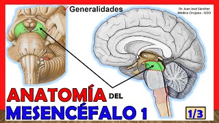
🥇 Anatomía del BULBO RAQUÍDEO. (Médula Oblongada) ¡Explicación Sencilla!
Introduction to the Anatomy of the Medulla Oblongata
In this video, José Sánchez introduces the structure of the medulla oblongata, also known as the bulbar region. The medulla oblongata is located at the posterior part of the brain and serves as a connection between the brain and spinal cord.
Structure of the Brain
- The brain can be divided into three main parts: anterior cerebrum (prosencephalon), middle brain (mesencephalon), and posterior brain (rhombencephalon).
- The medulla oblongata is part of the posterior brain and is further divided into two regions: metencephalon (superior portion) and myelencephalon (inferior portion).
Importance of the Medulla Oblongata
- The medulla oblongata is a crucial structure as it serves as a pathway for communication between the upper brain and spinal cord.
- It contains important nuclei responsible for various functions, including those associated with cranial nerves IX, X, XI, and XII.
- All ascending sensory fibers from the spinal cord pass through the medulla oblongata, while descending motor fibers from cortical or other encephalic regions also pass through it.
Anatomy of the Medulla Oblongata
- The medulla oblongata is located between the pons varolii (bridge) and spinal cord.
- It has a truncated pyramid shape with an anteriorly wider superior base.
- It passes through the foramen magnum but remains within the vertebral canal.
- There are two types of boundaries: a nervous boundary that is imprecise and an anatomical boundary that can be observed externally.
- The superior limit is defined by continuous sulcus limitans or pontomedullary sulcus.
- The inferior limit is more difficult to define precisely and is said to be immediately superior to the emergence of the anterior roots of the first cervical nerve.
Location and Boundaries of the Medulla Oblongata
In this section, José Sánchez explains the precise location and boundaries of the medulla oblongata.
Location within the Skull
- The medulla oblongata is located between the pons varolii (bridge) and spinal cord.
- It does not extend all the way down to the foramen magnum but remains within the vertebral canal.
Boundaries
- The superior boundary is defined by continuous sulcus limitans or pontomedullary sulcus.
- The inferior boundary is imprecise but is said to be immediately superior to the emergence of the anterior roots of the first cervical nerve.
Upper and Lower Limits of Medulla Oblongata
In this section, José Sánchez discusses in detail about the upper and lower limits of the medulla oblongata.
Upper Limit
- The upper limit of the medulla oblongata is well-defined by a continuous groove called sulcus limitans or pontomedullary sulcus.
- This groove contains important structures related to cranial nerves VI, VII, and VIII.
Lower Limit
- The lower limit of the medulla oblongata is imprecise and difficult to determine accurately.
- It is generally considered to be just above where anterior roots emerge from C1 spinal nerves.
- This lower limit coincides with decussation (crossing over) of pyramids, which will be discussed further in subsequent videos.
New Section
This section discusses the anatomical boundaries of the spinal cord and introduces the structures found in the anterior and lateral aspects of the medulla oblongata.
Anatomical Boundaries of the Spinal Cord
- The upper limit of the spinal cord is determined by a line that touches the anterior arch of the atlas (C1 vertebra) and the superior border of the posterior arch.
- Sometimes, this boundary may not correspond exactly to the actual anatomical limit due to variations in lamina placement.
- It is more accurate to refer to this boundary as a "nervous limit" rather than an anatomical one.
Structures in the Anterior and Lateral Aspects
- The anterior aspect of the medulla oblongata contains a median anterior groove or fissure, which continues upwards and terminates in a blind fossa called foramen cecum.
- The inferior part of this region includes decussation of pyramids, where fibers from the pyramidal tract cross over to opposite sides.
- Laterally, after crossing over from midline, we find two prominent structures: pyramids and olives.
- The pyramid is an elevation located lateral to the midline, while olives are bulges that push white matter outward.
- A groove called preolivary sulcus separates these structures. It gives rise to 10-12 fillets that form apparent origins of cranial nerve XII (hypoglossal nerve).
New Section
In this section, we continue exploring structures within the medulla oblongata, focusing on its anterior and lateral aspects.
Oliva Inferior and Preolivary Sulcus
- The inferior olive is an elevation located laterally next to pyramids. It pushes white matter outward, creating a prominence.
- The groove in front of the olive, separating it from the pyramid, is called preolivary sulcus or ventrolateral sulcus.
- This sulcus gives rise to 10-12 fillets that form the apparent origin of cranial nerve XII (hypoglossal nerve).
Postolivary Sulcus and Dorsolateral Sulcus
- Behind the olive, there is a groove called retroolivary sulcus. It creates a region known as the retroolivary zone.
- The dorsolateral sulcus, located further posteriorly, contains the apparent origins of cranial nerves IX (glossopharyngeal), X (vagus), and accessory.
New Section
This section continues discussing structures within the medulla oblongata, focusing on their locations and origins.
Retroolivary Sulcus vs. Dorsolateral Sulcus
- It is important to differentiate between the retroolivary sulcus and dorsolateral sulcus.
- The retroolivary sulcus is immediately behind the olive, while the dorsolateral sulcus is more posterior.
- The dorsolateral sulcus contains apparent origins of cranial nerves IX (glossopharyngeal), X (vagus), and accessory.
Origins of Cranial Nerves
- Some sources refer to these origins as being from the retroolivary sulcus, while others use the term dorsolateral sulcus.
- It's crucial to note that when studying live brain tissue, these grooves may not be visible as they appear in illustrations or diagrams.
New Section
In this section, we discuss variations in terminology regarding specific grooves within the medulla oblongata.
Terminology Variation
- There can be variation in the terminology used to describe specific grooves within the medulla oblongata.
- Some sources may refer to the dorsolateral sulcus as retroolivary sulcus, leading to confusion.
- It is important to be aware of these variations and understand that they may differ from what is commonly taught.
The transcript does not provide further information beyond this point.
New Section
This section discusses the origin of three cranial nerves and the anatomy of the medulla oblongata.
Origin of Cranial Nerves
- The retroolivary sulcus is where the cranial nerves emerge from the brainstem.
- The gloso-pharyngeal nerve (9th cranial nerve) originates superiorly, followed by the vagus nerve (10th cranial nerve), and then the accessory nerve (11th cranial nerve) which has its cranial root in the medulla oblongata.
- The medulla oblongata has a posterior half that forms part of the floor of the fourth ventricle and an inferior portion that lies outside this floor.
- The inferior portion contains the ependymal canal, which continues with the spinal cord's central canal.
Anatomy of Medulla Oblongata
- On the posterior surface of the medulla oblongata, there is a median sulcus similar to that found in the spinal cord. On either side of this sulcus are two prominences: medial tubercle gracilis and lateral tubercle cuneatus.
- The tubercle cuneatus gives rise to restiform body, which later forms inferior cerebellar peduncle connecting medulla oblongata to cerebellum.
- Other structures include intermediate posterior sulcus separating tubercle gracilis from tubercle cuneatus, dorsolateral sulcus seen on lateral aspect, and retroolivary sulcus where spinal roots of accessory nerve emerge.
New Section
This section explores further details about specific structures in relation to their origins and connections within the brainstem.
Trigeminal Tubercle and Inferior Cerebellar Peduncle
- The trigeminal tubercle is an elevation in the medulla oblongata, specifically in the medullary nucleus of the trigeminal nerve.
- The inferior cerebellar peduncle originates from the tubercle cuneatus and extends upwards, forming a connection between the medulla oblongata and cerebellum.
Additional Structures
- Other structures include the lateral sulcus, retroolivary sulcus (which is not related to the medulla oblongata), and preolivary sulcus.
- The preolivary sulcus is where the spinal portion of the accessory nerve emerges.
- The hypoglossal nerve (12th cranial nerve) originates from this region as well.
Please refer to the video for a more detailed explanation and visual representation of these structures.
Turn any video into a summary like this
YouTube links, meetings, lectures. With transcripts, search, and chat.