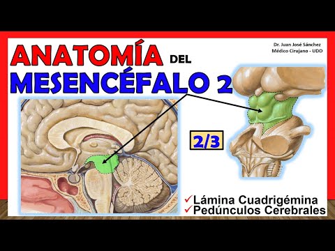
🥇 Anatomía del MESENCÉFALO 2/3. (Pedúnculos Cerebrales, Lámina Cuadrigémina). ¡Facil Explicación!
Introduction to the Mesencephalon
Overview of the Mesencephalon
- The video introduces the mesencephalon, also known as the midbrain, as part of a series on anatomical structures.
- It outlines that this is the second of thirteen installments focusing on different aspects of the mesencephalon.
Structure and Components
- The discussion will cover two main parts: the quadrigeminal lamina and cerebral peduncles, with a future video dedicated to the aqueduct of Sylvius.
- The brain is divided into three sections: forebrain (prosencephalon), midbrain (mesencephalon), and hindbrain (rhombencephalon).
Detailed Anatomy of the Mesencephalon
Subdivisions and Terminology
- The mesencephalon does not have subdivisions like other brain regions; it consists mainly of a green-highlighted area in diagrams.
- The term "mesencephalon" derives from Greek, meaning "middle," indicating its position in relation to other brain structures.
Cerebral Peduncles
- The cerebral peduncles are described as stalk-like structures that connect various parts of the brain, particularly linking to the cerebellum.
- They appear to emerge from below and integrate with cerebral hemispheres, specifically involving subthalamic areas and internal capsule regions.
Functional Aspects of Cerebral Peduncles
Importance in Brain Structure
- It's crucial to refer to these structures as "cerebral peduncles" for clarity since there are three distinct types identified.
- A sagittal cut reveals an anterior region (cerebral peduncles) and a posterior region called tectal or quadrigeminal lamina.
Recap on Mesencephalonic Structure
- In summary, the mesencephalon comprises an anterior portion (cerebral peduncles) and a posterior section (tectal lamina).
- The anterior segment includes specific features like substantia nigra while posterior segments relate closely to visual processing centers.
Exploring Tectal Lamina Structures
Quadrigeminal Lamina Composition
- The tectal lamina is formed by four colliculi which play significant roles in sensory processing.
Conclusion & Call-to-action
Overview of the Mesencephalon Structure
Introduction to the Mesencephalon
- The discussion begins with a focus on the mesencephalon, specifically its anterior region and the development of the tectal plate.
- The term "cruciform sulcus" is introduced, describing a longitudinal line that separates colliculi into upper and lower sections.
Superior and Inferior Colliculi
- The superior colliculi are noted for being larger than inferior ones, housing the pineal gland in their central groove.
- Superior colliculi connect laterally via white matter structures known as the superior colliculus arm, which interacts with the lateral geniculate body related to visual reflexes.
- In contrast, inferior colliculi are smaller and positioned more medially; they connect through a structure called the inferior colliculus arm to auditory pathways.
Structural Features of Mesencephalon
- A cross-sectional image illustrates key features of the mesencephalon, including its anterior and posterior aspects.
- The peduncles appear separated from an anterior view but converge posteriorly to form a significant segment of mesencephalic structure.
Surfaces of the Mesencephalon
- The junction where peduncles meet forms a lateral sulcus in Portuguese terminology; this is crucial for understanding mesencephalic anatomy.
- Four surfaces are identified: anteroinferior, posteroinferior, lateral (one on each side), and medial (central).
Anteroinferior Surface Details
- The anteroinferior surface is convex and formed by parts of cerebral peduncles; it also includes elements like interpeduncular fossa.
- This fossa has specific boundaries: anteriorly by optic chiasm, laterally by optic tracts, medially by peduncle borders, and posteriorly by mesencephalic sulcus.
Posterior Superior Surface Insights
- The posterior superior surface is defined by tectal laminae; it connects superior cerebellar peduncles with functions related to cerebellar coordination.
Mesencephalon Overview
Key Structures and Features
- The frenulum and superior medullary velum connect structures in the mesencephalon, marking the emergence of the fourth cranial nerve (trochlear nerve) from its apparent origin.
- The lateral sulcus of the mesencephalon is highlighted; it is crucial to remember this landmark as it plays a significant role in neuroanatomy.
- The lateral sulcus extends from the junction of the superior cerebellar peduncle to the inferior colliculus, providing a clear demarcation within the mesencephalon.
- This marked sulcus serves as a reference line for identifying important structures such as the lateral trigone, which is defined by its base at this sulcus and apex at the inferior colliculus.
- Understanding this anatomy is essential for recognizing pathways in neuroanatomy, particularly when studying neurological functions related to these structures.
Medial Aspects and Cranial Nerves
- The medial aspect of both cerebral peduncles reveals part of the oculomotor space and highlights where the third cranial nerve (oculomotor nerve) emerges.
- This anatomical knowledge aids in understanding how various neural pathways interact within the brainstem, especially concerning motor control and eye movement.