🥇 1er Par Craneal - NERVIO OLFATORIO (1 de 2). Origen Real y Aparente, Trayecto y Relaciones
Introduction to the Olfactory Nerve
Overview of Cranial Nerves
- The video introduces the topic of cranial nerves, specifically focusing on the first cranial nerve, known as the olfactory nerve.
- It mentions that there will be two videos dedicated to this nerve, starting with its origin and pathway.
Understanding Cranial Nerves
- There are traditionally 12 pairs of cranial nerves, with a mention of a 13th pair (the terminal nerve), which will be discussed separately.
- The olfactory and optic nerves are considered extensions of the forebrain (prosencephalon).
Olfactory Nerve Characteristics
Origin and Pathway
- The concept of "real origin" vs. "apparent origin" is introduced; apparent origin refers to where the nerve appears to emerge from the brain.
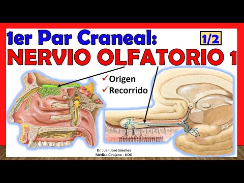
- The apparent origin for the olfactory nerve is at the base of the olfactory bulb.
Naming Convention
- Cranial nerves are internationally designated using Roman numerals; thus, the olfactory nerve is referred to as I.
Real Origin and Unique Features
Real Origin Explained
- The real origin involves bipolar cells located in the olfactory mucosa, which serve as sensory receptors for smell.
- Unlike other cranial nerves, both olfactory and optic nerves do not have nuclei in the brainstem.
Pathway from Reception to Brain
Receptor Apparatus
- Discussion shifts towards how information is received by receptor cells located in nasal mucosa.
- Two regions within nasal cavity: lateral (where turbinates are located) and medial (nasal septum).
Cellular Structure
- Bipolar cells or "olfactory receptor neurons" play a crucial role in detecting odor stimuli through their cilia.
Impact on Sensory Functionality
Effects on Olfaction
- Conditions affecting nasal epithelium can lead to diminished sense of smell due to impaired function of cilia on bipolar cells.
Formation of Olfactory Nerve
Olfactory System Overview
Structure of the Olfactory Nerve
- The olfactory system begins with the first neurons of the olfactory nerve, which consist of approximately 20 filaments from each nasal cavity.
- These filaments are made up of hundreds of thousands to millions of bipolar olfactory cells, with each filament containing around 20,000 bipolar cells.
Pathway and Arrangement
- The axons travel superiorly through the cribriform plate of the ethmoid bone, arranged in two groups: medial and lateral.
- An analogy is drawn comparing these arrangements to a toothbrush, where vertical and oblique filaments represent different orientations.
Bulb Structure and Function
- The olfactory nerve traverses the cribriform plate to reach the inferior surface of the olfactory bulb, which resembles a toothbrush head.
- Surrounding structures include a layer of pia mater before reaching the olfactory bulb; dura mater is also present but does not envelop the nerve directly.
Importance of Subscribing
- Viewers are encouraged to subscribe for more anatomical videos and share their university or country in comments for engagement.
Understanding Olfactory Bulbs
Location and Anatomy
- Each side has an olfactory bulb located above the cribriform plate in the anterior cranial fossa, corresponding to each nasal cavity.
- The bulbs have an ovoid shape that is flattened anteroposteriorly and elongated laterally.
Relationship with Brain Structures
- The frontal lobe's inferior surface features a groove known as the olfactory sulcus that corresponds with both bulbs and their respective tracts.
Cellular Composition
- Axons from bipolar cells enter the bulbus; these are crucial for understanding how signals are processed within this structure.
Cell Types in Olfactory Bulb
Key Cell Types Identified
- Four main cell types exist within the bulbus: mitral cells, tufted (or penacho) cells, granule cells, and glomerular cells.
- Mitral and tufted cells serve as primary relay neurons in processing sensory information from bipolar cell axons entering into them.
Understanding Olfactory Processing
The Role of Bipolar Cells and Glomeruli
- The connection between bipolar cells and mitral or tufted cells is referred to as a glomerulus, marking the first synapse in the olfactory system.
- Each glomerulus can receive approximately 25,000 axonal connections from bipolar cells, indicating a high level of convergence in olfactory stimuli processing.
- Each glomerulus is specific to particular olfactory information, allowing for diverse stimuli to converge into fewer outputs.
- Axons from mitral and tufted cells exit the olfactory bulb to form the olfactory tract, which transmits impulses to cortical pathways.
Integration and Processing within the Olfactory Bulb
- Granule and periglomerular cells play crucial roles in processing and integrating information within the olfactory bulb rather than receiving sensory input directly.
- These interneurons interconnect with mitral and tufted cells, facilitating integration of olfactory signals through feedback mechanisms.
- Interneurons receive contralateral information from other olfactory bulbs, enhancing integrative processing capabilities.
Structure of the Olfactory Tract
- The olfactory tract extends 3 to 3.5 centimeters from the bulb towards deeper brain structures, specifically targeting areas associated with smell perception.
- It bifurcates into medial and lateral olfactory striae at its posterior end near the anterior perforated substance.
Key Centers in Olfactory Processing
- The anterior olfactory nucleus is located within peripheral gray matter along the tract; it serves as one of the initial centers for processing incoming signals.
- Three types of synapses occur at this central white matter: connections leading to the anterior nucleus, long axonal projections to cortical areas, and links to other significant structures.
Understanding Olfactory Striae Functionality
- The medial stria connects more centrally while lateral stria are larger and extend further laterally toward amygdala-related regions for emotional context in smell perception.
Olfactory System: Understanding the Medial Olfactory Stria
Structure and Function of the Medial Olfactory Stria
- The medial olfactory stria is described as thinner and shorter compared to the lateral stria, terminating at the medial surface of the frontal lobe.
- Visual representation in diagrams helps illustrate the differences between medial and lateral olfactory striae, emphasizing their distinct characteristics.
Broca's Olfactory Junction
- The diagonal band forms what is known as Broca's olfactory junction, a significant area for understanding olfactory pathways.