Los OJOS: cómo funcionan, anatomía, partes, funciones, sentido de la vista
Understanding the Complexity of Human Eyes
The Role and Function of Eyes
- The eyes are essential organs that enable us to perceive our surroundings, forming one of the five senses alongside touch, hearing, smell, and taste. Vision is a complex process involving various parts of the eye.
- Eyes serve as mobile cameras located at the front of our face, allowing us to gather visual information about colors, shapes, distances, sizes, and spatial depth.
- The vision process begins when light reflects off objects and enters the eyes. This light is converted into chemical or electrical signals that the brain interprets as images.
Anatomy of the Eye
- Each human has two eyes positioned in hollow cavities known as ocular orbits. These orbits are shaped like pyramids with specific bones forming their structure: frontal, sphenoid, zygomatic, maxillary, ethmoid, lacrimal, and palatine.
- Extraocular muscles connect to each eye within these orbits. They allow for movement in multiple directions and are categorized into two groups: rectus (four muscles) and oblique (two muscles).
Protective Features of Eyes
- Eyelids protect the eyes from excessive light and foreign particles while keeping them moist through blinking—a combination of voluntary and involuntary action.
- The conjunctiva is a transparent mucous membrane covering both eyelids' inner surfaces and the eyeball's surface. It protects against infections and aids in tear production.
Tear Production Mechanism
- Tears consist of three components: aqueous (watery), oily (lipid), and mucous layers. Aqueous tears are produced by lacrimal glands located beneath the eyebrows; oily tears come from Meibomian glands in eyelids; mucous tears originate from conjunctival membranes.
Structural Components of the Eye
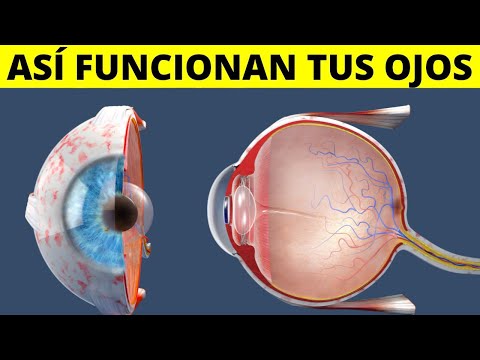
- The sclera is the white part of the eye made from tough tissue that covers most of it. It contains blood vessels supplying nutrients to ocular cells.
- In front of the colored part lies a transparent dome called cornea which helps focus incoming light onto objects we observe.
Understanding the Human Eye
Anatomy of the Eye
- The iris, which is the colored part of our eyes, is located just behind the cornea and plays a crucial role in regulating light entry.
- The pupil, appearing as a black dot in the center of the iris, adjusts size based on light intensity due to muscle contractions in the iris.
- In bright environments, the pupil constricts to limit light intake; conversely, it dilates in darkness to allow more light for better vision.
Lens and Retina Functionality
- The lens (or crystalline lens), situated behind the iris, focuses light onto the retina and changes shape for near or distant vision.
- The retina contains millions of light-sensitive cells that convert light into nerve impulses sent to the brain for visual interpretation.
Types of Photoreceptor Cells
- There are two types of photoreceptors: rods (for black-and-white vision and shapes) and cones (for color detection).
- The macula within the retina is responsible for central vision and detail recognition.
Supporting Structures
- The choroid membrane lies between the sclera and retina; it nourishes retinal cells and reflects light, causing red-eye effects in photos.