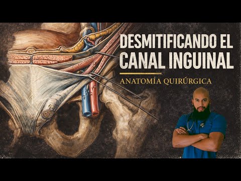
ANATOMÍA - CANAL INGUINAL ACTUALIZADO (Dependencias de las Fascias, Epónimos, Límites & Anillos)
Anatomía del Canal Inguinal: Desmitificando Conceptos
Introducción a la Anatomía del Canal Inguinal
- El presentador introduce el tema de la anatomía del canal inguinal desde una perspectiva quirúrgica, mencionando que es una actualización de un video anterior.
- Se destaca que la región inguinal es un área compleja entre el abdomen y el muslo, donde estructuras vasculonerviosas pasan hacia el miembro inferior.
Controversias en Terminología Anatómica
- Se menciona que hay discrepancias en la terminología utilizada para describir las estructuras del canal inguinal, tanto en artículos científicos como en libros clásicos.
- El presentador se propone desmitificar estas controversias y aclarar conceptos clave sobre el canal inguinal.
Definición: ¿Canal o Conducto?
- Se explica que un "conducto" tiene paredes propias, mientras que el "canal inguinal" es un túnel sin paredes definidas, dependiente de otras estructuras musculares.
- La longitud promedio del canal inguinal en adultos es de aproximadamente 4 cm.
Importancia de la Embriología
- Para entender bien la anatomía del canal inguinal, es crucial conocer su embriología; se menciona el descenso testicular como evento clave durante la séptima semana de vida intrauterina.
- Los testículos inicialmente están en la región lumbar y descienden guiados por una estructura llamada gubernáculo.
Estructuras Asociadas al Canal Inguinal
- El contenido del canal inguinal incluye el cordón espermático en hombres y el ligamento redondo del útero en mujeres.
- Es fundamental conocer las formaciones dependientes de las aponeurosis y fascias abdominales para evitar confusiones terminológicas.
Anatomy of the Inguinal Ligament and Related Structures
Overview of the Inguinal Ligament
- The inguinal ligament is known by several names, including Colles' ligament, reflex inguinal ligament, and posterior pillar of the external oblique. Each term will be defined and explored in detail.
- It is crucial to clarify that "inguinal" can refer to different structures, leading to confusion; emphasis will be placed on these distinctions.
Anatomical Features
- The inguinal ligament originates from the anterior superior iliac spine (ASIS) and inserts at the pubic tubercle. This anatomical pathway is a classic question in anatomy studies.
- Important muscular and vascular structures pass beneath the inguinal ligament, including the iliacus muscle, pectineus muscle, femoral vessels, and femoral nerve. These relationships will be elaborated upon in subsequent discussions.
Structural Relationships
- The image presented illustrates how the aponeurosis of the external oblique muscle forms the inguinal ligament; this visual aid helps clarify its structure.
- A notable feature is a deflection or dependency from the inguinal ligament called the lacunar ligament; this relationship will be further explained later on. Additionally, an important structure called the iliopectineal arch is located beneath it.
Understanding Related Structures
- The iliopubic tract runs parallel and deep to the inguinal ligament but can only be observed from a posterior view; it follows a similar trajectory as that of the inguinal ligament itself.
- The iliopubic tract represents a thickening of transversalis fascia that also extends between ASIS and pubic spine, providing structural support within this region.
Division of Compartments Underneath Inguinal Ligament
- The iliopectineal arch serves as a division between two compartments:
- Lateral Compartment (Muscular): Contains structures such as femoral nerve and iliacus muscle.
- Medial Compartment (Vascular): Known as vascular lagoon where key elements like femoral vessels are found alongside lymph nodes referred to as deep inguinal lymph nodes or historically named Clock's node or Rosenmüller's node. This distinction is vital for anatomical understanding.
Anatomical Structures in the Inguinal Region
The Femoral Ring and Lacunar Ligament
- The femoral ring is a small anatomical structure located within the vascular lacuna, positioned medial to the femoral vein.
- The lacunar ligament originates from the inguinal ligament and extends downward, terminating at a bony prominence known as the pecten of the pubis.
- The lacunar ligament serves as the medial boundary of the femoral ring, highlighting its significance in anatomical orientation.
Pectineal Ligament
- Identified as one of the strongest ligaments in this region, the pectineal ligament is an important reinforcement structure.
- It is essentially a thickening of the periosteum of the coxal bone and receives additional support from both the lacunar ligament and other surrounding structures.
- The pectineal ligament is reinforced by fibers from adjacent fasciae, including those associated with the pectineus muscle.
Interfobiolar Ligament (Gesselbach's Ligament)
- This ligament is an extension of transversalis fascia that begins at a specific point known as Douglas' line or arcuate line.
- It follows an arc-like path downwards towards its insertion on the hilopubic tract, playing a crucial role in reinforcing abdominal wall integrity.
- Positioned medially to the deep inguinal ring, it contributes significantly to posterior canal support.
Conjoint Tendon
- The conjoint tendon represents a fusion between internal oblique aponeurosis and transverse muscle fibers, providing structural reinforcement to abdominal walls.
- Its insertion occurs at the pubic tubercle; however, it can be variable in presence among individuals.
This structured overview captures key anatomical concepts discussed within specified timestamps while maintaining clarity for study purposes.
Understanding the Conjoined Tendon and Inguinal Structures
Overview of the Conjoined Tendon
- The conjoined tendon is present in approximately 3-5% of the population, making it relatively rare. It is formed by the fusion of the aponeurosis of the internal oblique muscle and the transversus abdominis muscle, inserting at the pubic tubercle.
Definition and Confusion with Oinguinal
- The term "oz inguinal" refers to what was historically known as Henle's ligament, which is distinct from the conjoined tendon but often confused due to their similar insertion points and functions. Both structures reinforce the posterior wall of the inguinal canal.
Differences Between Structures
- While both structures insert at similar locations, their origins differ:
- The conjoined tendon arises from a fusion of specific abdominal muscles.
- The oz inguinal (Henle's ligament) originates from a lateral pillar associated with the rectus abdominis muscle.
- Oz inguinal is more common, found in about 30-50% of individuals.
Terminology Clarification
- Recent anatomical literature suggests using a unified term "área conjunta" to refer collectively to both structures (oz inguinal and conjoined tendon), addressing historical terminological ambiguities. This reflects contemporary understanding in anatomy studies.
Fascia Transversalis Explained
- The fascia transversalis or endoabdominal fascia is a connective tissue layer that lines the inner abdomen, separating muscles from peritoneal fat; it extends superiorly to reach the diaphragm and laterally connects with other abdominal structures while anchoring inferiorly at key pelvic landmarks like iliac crests.
Anatomy of the Inguinal Canal
Structure of Inguinal Canal
- The inguinal canal has a quadrangular prism shape with four walls: anterior, posterior, superior (roof), and inferior (floor). Each wall plays a crucial role in its structural integrity and function within abdominal anatomy.
Anterior Wall Composition
- Formed primarily by the aponeurosis of external oblique muscle throughout its length; reinforced laterally by muscular fibers from internal oblique muscle for added support against potential hernias.
Superior Wall Characteristics
- Composed mainly of fibers from both internal oblique and transversus abdominis muscles; this roof structure provides critical support for contents passing through or residing within the canal area.
Posterior Wall Importance
- Considered vital surgically as it represents an area where hernias can protrude; constructed by deep layers including transversus abdominis fascia and endoabdominal fascia which provide necessary reinforcement against such occurrences.
Anatomical Details of the Inguinal Canal
Overview of the Posterior Wall
- The posterior wall is formed by the transversalis fascia, which has important anatomical details. The medial third is reinforced by the conjoint tendon.
Medial and Lateral Third Reinforcements
- The medial third is supported by the conjoint tendon, while the lateral third is reinforced by the interfobiolar ligament. This creates a vulnerable area in between.
Hesselbach's Triangle
- A small triangle known as Hesselbach's triangle, located in the middle third, lacks reinforcement and is where direct inguinal hernias can protrude.
Formation of the Floor
- The floor of the inguinal canal consists of two structures:
- Anteriorly formed by the inguinal ligament.
- Posteriorly formed by Thomson's iliopubic tract (iliopubian band).
Summary of Canal Structure
- Key components include:
- Anterior wall: external oblique aponeurosis.
- Roof: internal oblique and transversus abdominis forming conjoint tendon.
- Posterior wall: transversalis fascia with interfobiolar ligament.
- Floor: inguinal ligament anteriorly and iliopubic tract posteriorly.
Understanding Inguinal Rings
Description of Inguinal Rings
- The inguinal canal contains two openings:
- Deep inguinal ring (entrance for spermatic cord).
- Superficial inguinal ring (exit point).
Terminology Clarification
- Historically, deep inguinal ring was referred to as "internal," causing confusion; it’s now called "deep" to clarify its position relative to abdominal cavity. Similarly, superficial ring was previously termed "external."
Location and Identification
- The deep inguinal ring appears as an oval opening in transversalis fascia, located about 1 cm above midpoint of inguinal ligament. It should not be confused with other structures below it.
Weakness Areas in Hesselbach's Triangle
Characteristics of Hesselbach's Triangle
- Defined boundaries include:
- Rectus abdominis muscle,
- Epigastric vessels,
- Inguinal ligament with iliopubic tract below.
This area represents a weakness due to lack of muscular protection; only covered by fused aponeuroses from internal oblique and transversus abdominis muscles.
Superficial Inguinal Ring Anatomy
Structure Description
- The superficial inguinal ring features well-defined borders:
- Medial pillar inserts into pubic crest from external oblique muscle.
- Lateral pillar attaches at pubic tubercle also from external oblique muscle.
- Both pillars are connected via arcuate fibers known as intercolumnar or arciform fibers.
Anatomy of the Inguinal Region
Ligaments and Structures in the Inguinal Area
- The intercolumnar or arched ligaments connect the medial and lateral pillars, with a notable mention of the posterior pillar of the external oblique, known as Colles' ligament.
- The posterior pillar is crucial for understanding the superficial inguinal ring's anatomy; it consists of three pillars: medial, lateral, and posterior.
Myopectineal Orifice
- The myopectineal orifice of Fruchaud is an area within the abdomen that represents a fusion zone where abdominal wall weaknesses can lead to inguinal and femoral hernias.
- This orifice has specific boundaries:
- Superiorly defined by the inferior border of the transversus abdominis muscle.
- Laterally bounded by the iliopsoas muscle.
- Medially marked by the rectus abdominis muscle.
- Inferiorly limited by the superior pubic ramus.
Hernia Types and Pathways
- The myopectineal area is divided into two planes:
- An upper plane for direct and indirect inguinal hernias (indicated as A for indirect through deep inguinal ring, B for direct through Hesselbach's triangle).
- A lower plane designated for femoral hernias.
- Structures traversing this opening include:
- Spermatic cord in males (round ligament in females).
- Femoral vessels (femoral artery and vein).
- The internal aspect of this opening is primarily closed off by transversalis fascia.