Radioterapia Aula 04
Introduction to Music and Equipment Evolution
In this section, the speaker briefly discusses concepts related to music, equipment, and the evolution of techniques in radiotherapy.
Concepts Discussed:
- The importance of accelerators in radiotherapy.
- The introduction of special X-ray equipment and telecobalt accelerators.
- The benefits of improved equipment and accelerator technology in radiation treatment.
Advancements in Techniques and Positioning
This section focuses on advancements in techniques and positioning in radiotherapy.
Key Points:
- The use of 2D conventional techniques versus 3D techniques.
- In 2D techniques, treatment planning was based on two-dimensional images.
- With the introduction of 3D technology, volumetric definition of structures became possible.
- Individualized techniques and personalized dose calculations became feasible with 3D technology.
Benefits of 3D Techniques
This section highlights the benefits of using 3D techniques in radiotherapy.
Key Points:
- Precise volume definition and well-defined margins for treatment planning.
- Improved control over tumor growth and eradication.
- Individualized treatment plans based on patient-specific parameters.
- Enhanced accuracy in targeting tumors while minimizing risks to surrounding organs.
Individualized Treatment Planning
This section discusses individualized treatment planning in radiotherapy.
Key Points:
- Different areas of medicine, such as nuclear medicine, require individualized treatment planning.
- Each patient has a specific treatment plan tailored to their needs and calculated doses.
- Individualized planning allows for greater precision in targeting medical targets.
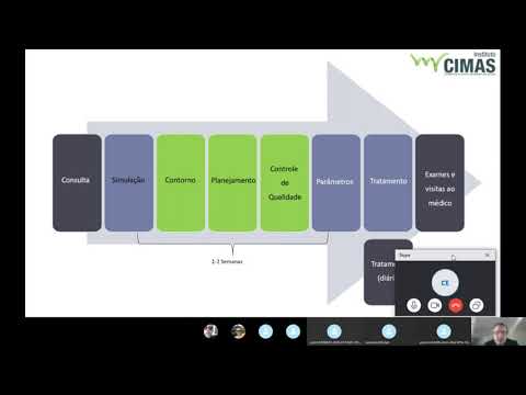
Steps in the Radiation Treatment Process
This section outlines the various steps involved in radiation treatment.
Key Points:
- The process begins with a consultation and medical history review.
- Simulation and imaging are performed to aid in treatment planning.
- Contouring and quality control measures are implemented for accurate treatment delivery.
- The duration of the treatment process can vary from one to two weeks, depending on factors such as hospital availability.
Advancements in Technology for Faster Processes
This section discusses advancements in technology that have accelerated certain aspects of the radiation treatment process.
Key Points:
- Multileaf collimation technology allows for faster contouring and mold creation.
- Previously, molds were manually created, which was time-consuming.
- Advances in technology have reduced the time required for mold creation and quality control processes.
Challenges in Implementing Multileaf Collimation
This section addresses challenges faced by hospitals during the implementation of multileaf collimation technology.
Key Points:
- Some hospitals faced difficulties due to financial constraints and software updates.
- Acquiring equipment compatible with multileaf collimators took time for some institutions.
- Migration to multileaf collimation required adjustments and investments.
Parameters Analysis and Treatment Schedule
This section focuses on analyzing parameters related to radiation treatment and discussing the treatment schedule.
Key Points:
- Parameters analysis is crucial for effective treatment planning.
- Treatment typically involves daily doses administered five times a week (Monday to Friday).
- Regular follow-up visits are scheduled for monitoring progress or additional treatments if needed.
Consultation Process and Boost Treatments
This section explains the consultation process and the possibility of boost treatments.
Key Points:
- The initial consultation involves diagnosing and measuring the tumor, reviewing medical history, and conducting physical examinations.
- Boost treatments may be required for additional radiation doses.
- The number of boost sessions depends on the prescribed dose and individual patient factors.
The transcript is in Portuguese.
Introduction to Treatment Planning
In this section, the speaker discusses the distribution of treatment sessions and the importance of radiobiology in treatment planning.
Distribution of Treatment Sessions
- The speaker mentions that there will be around 53 treatment sessions.
- The distribution of these sessions is determined by considering factors such as tumor control probability and normal tissue complication probability.
Importance of Radiobiology in Treatment Planning
- Radiobiology provides parameters for individualized treatment schedules based on the probability of tumor control and normal tissue complications.
- The total dose and number of fractions are also based on radiobiological considerations.
- A dose of 50 Greys is sufficient to eradicate tumor cells, but giving a higher dose may lead to more side effects and potential harm to healthy tissues.
- The time required for the entire treatment is also taken into account during treatment planning.
Radiobiology Considerations in Treatment Planning
This section focuses on the role of radiobiology in determining the appropriate treatment for patients.
Fractionation and Dose Redistribution
- Fractionation involves dividing the total radiation dose into smaller daily doses.
- This approach aims to minimize toxicity while maintaining effectiveness in treating tumors.
- The concept of redistribution refers to targeting cells at their most sensitive phases during fractionated treatments.
- Repopulation, which refers to cell growth after radiotherapy, is also considered when determining the duration and timing of treatments.
Five Rs of Radiobiology
- Repair: Cells respond differently to radiation depending on whether they are tumor cells or healthy cells. Tumor cells with a higher mitotic rate freeze during checkpoint cell cycle repair, while healthy cells recover faster from radiation damage.
- Repopulation: Some cells escape radiation-induced death and continue to grow. These repopulating cells can be targeted at specific points during treatment.
- Redistribution: The sensitivity of cells to radiation varies depending on their cell cycle phase. Cells in the G2 and mitotic phases are more radiosensitive, making fractionation an effective strategy.
- Reoxygenation: Oxygen plays a crucial role in enhancing the effectiveness of radiation therapy. Tumor cells in well-oxygenated areas are more susceptible to radiation damage.
- Radiosensitivity: Different types of cells have varying levels of sensitivity to radiation, with tumor cells often being more radiosensitive than healthy cells.
Repopulation and Treatment Duration
This section discusses repopulation as a factor in treatment planning and the importance of adhering to the recommended treatment duration.
Repopulation
- Repopulation refers to the growth of cells that survive radiotherapy.
- Some cells may escape radiation-induced death due to variations in their cell cycle phases.
- These surviving cells continue to multiply, leading to repopulation within the treated area.
Importance of Treatment Duration
- The duration of treatment should be carefully planned and adhered to.
- By following the recommended treatment duration, it is ensured that all necessary doses are delivered within an appropriate timeframe.
- Adhering to the treatment schedule helps maximize effectiveness while minimizing potential harm to healthy tissues.
The transcript provided does not cover all aspects discussed in the video.
Reoxygenation and Radiosensitivity
This section discusses the importance of oxygen in cells and its role in repairing radiation damage. It also explains how radiation treatment can target hypoxic cells and the concept of radiosensitivity.
Reoxygenation and Damage Fixation
- Oxygen is essential for repairing damage in cells caused by radiation.
- Approximately 30% of cells are hypoxic, meaning they lack oxygen.
- When irradiated, these hypoxic cells are gradually eliminated, allowing revascularization to occur.
- Revascularization brings oxygen to previously hypoxic areas, enabling damage fixation.
Fractionation and Radiosensitivity
- Fractionation refers to the gradual killing of cells during radiation treatment.
- Some cells are more radiosensitive, while others are more radioprotective.
- Treatment must be tailored to respect the tumor control sensitivity of different cell types.
- Dose total and fractionation are determined based on specific pathologies.
Repopulation and Radiobiology
This section explores repopulation as a factor in radiation treatment. It also discusses the concept of radiobiology and how different organs have varying tolerances to radiation.
Repopulation
- Repopulation refers to the regrowth of surviving tumor cells during treatment intervals.
- The rate of repopulation affects the overall duration of treatment.
Radiobiology and Tumor Control
- Different organs have varying sensitivities to radiation.
- Treatment planning considers both tumor control sensitivity and normal tissue tolerance levels.
- Overdosing or underdosing can impact treatment efficacy.
Simulation and Contouring
This section focuses on simulation and contouring in radiotherapy planning. It explains the importance of accurate target localization, risk organ identification, and the use of accessories and immobilizers.
Simulation and Contouring
- Simulation involves acquiring images, such as CT scans, for treatment planning.
- The physician is responsible for contouring and defining target volumes and risk organs.
- Accessories and immobilizers are crucial for maintaining reproducibility throughout treatment.
Challenges in Radiotherapy Planning
This section highlights the challenges faced during radiotherapy planning, including individualized treatment, variability in patient conditions, and the use of advanced technologies to aid in planning.
Individualized Treatment
- Each patient requires a personalized treatment plan based on their specific condition.
- Treatment cannot be standardized or reused between patients with similar diseases.
Variability in Patient Conditions
- Factors such as diagnosis, tumor size, and field size influence treatment planning.
- Reproducibility is essential but can be challenging due to various patient-specific factors.
Role of Advanced Technologies
- New technologies aid in accurate target delineation and visualization during planning.
- Reconstruction methods within software assist in precise contouring of structures.
Importance of Imaging Technologies
This section emphasizes the significance of imaging technologies in radiotherapy planning. It discusses how new technologies improve visualization, aid in target identification, and enhance overall treatment planning.
Imaging Technologies
- Advanced imaging techniques play a crucial role in radiotherapy planning.
- PET scans combined with other imaging modalities help identify metabolic activity within tumors.
- Accurate visualization assists physicians in determining appropriate treatment parameters.
Desenho dos Volumes
This section discusses the importance of accurately defining volumes in radiation treatment planning. The different volumes necessary for planning are explained, including GTV (Gross Tumor Volume), CTV (Clinical Target Volume), ITV (Internal Target Volume), PTV (Planning Target Volume), and OAR (Organs at Risk).
Definition of Volumes
- GTV is the volume to be treated and is determined based on physical examination and relevant investigations.
- Other volumes include CTV, ITV, PTV, and OAR.
GTV (Gross Tumor Volume)
- GTV is defined by physical examination and relevant investigations.
- It represents the tumor or disease that needs to be eradicated.
CTV (Clinical Target Volume)
- CTV is a margin around the GTV that accounts for microscopic neoplasia or areas that may have been missed during contouring.
- Margins for CTV depend on various factors such as external images, patient parameters, alignment, organ movement, etc.
ITV (Internal Target Volume)
- ITV considers internal margins related to bladder filling, respiration, etc.
- It ensures that the target remains within the radiation field during treatment.
PTV (Planning Target Volume)
- PTV includes additional margins beyond ITV to account for uncertainties in patient setup and treatment delivery.
- Reproducibility of patient positioning and alignment are important considerations for PTV margins.
OAR (Organs at Risk)
- OAR refers to organs near the target volume that need to be spared from excessive radiation dose.
- Accurate delineation of OAR helps in minimizing potential side effects.
Considerations in Treatment Planning
This section discusses various considerations in treatment planning. Factors such as patient parameters, organ movement, weight loss, and alignment are important for accurate treatment delivery.
Margins and Considerations
- Margins for target volumes depend on factors like patient parameters, external images, organ movement, etc.
- Internal margins account for bladder filling, respiration, and other internal factors.
- Reproducibility of patient positioning and alignment is crucial for maintaining consistent margins.
Patient Parameters
- Changes in patient weight or organ size can affect the accuracy of treatment planning.
- Regular monitoring and adjustments may be necessary to ensure proper targeting.
Alignment
- Proper alignment of the patient during treatment is essential to maintain accurate targeting.
- The use of immobilization devices helps in reproducibility and minimizing setup errors.
Simulation Process
This section explains the simulation process involved in radiation treatment planning. It includes considerations such as patient preparation, imaging modalities, and maintaining consistency between planning and treatment sessions.
Simulation Process
- Patient preparation involves following specific instructions regarding bladder and bowel emptying.
- Imaging modalities like CT scans provide detailed information for contouring target volumes.
- Consistency between planning and treatment sessions is important to ensure accurate delivery of radiation.
Conclusion
This section concludes the discussion on volume delineation in radiation treatment planning. Accurate definition of volumes is crucial for effective treatment while considering various factors that may impact target coverage.
Importance of Volume Delineation
- Accurate volume delineation ensures effective targeting of tumor or disease.
- Considerations such as organ movement, patient parameters, weight loss, etc., should be taken into account during volume delineation.
Timestamps have been associated with relevant bullet points to facilitate easy navigation through the transcript.
Understanding Radiotherapy Planning
In this section, the speaker discusses the importance of considering organ movement in radiotherapy planning. They explain the concept of GTV (Gross Tumor Volume), CTV (Clinical Target Volume), and PTV (Planning Target Volume). The speaker also mentions the significance of identifying and protecting organs at risk during treatment.
GTV, CTV, and PTV
- GTV refers to the volume of the tumor that needs to be treated.
- CTV is a margin around the GTV that accounts for microscopic disease or areas not visible to the naked eye.
- PTV is the planning target volume that considers both external and internal parameters for accurate dose calculation.
Organs at Risk
- Organs at risk are organs with low tolerance to radiation.
- Examples of organs at risk include the brain, spinal cord, parotid glands, and optic nerves.
- Efforts are made to avoid irradiating these organs as much as possible during treatment.
Balancing Risks and Benefits
- Avoiding organs at risk entirely may not always be feasible due to individual patient circumstances.
- A cost-benefit analysis is conducted in such cases, involving discussions between medical professionals and patients.
Importance of Patient Positioning and Immobilization
This section emphasizes the significance of precise patient positioning and immobilization in radiotherapy. The speaker explains how accurate positioning allows for reproducibility in treatment delivery.
Precise Positioning
- Accurate patient positioning is crucial for effective radiotherapy.
- Proper immobilization and support devices, such as headrests, are used to ensure precise positioning.
- Correct positioning allows for millimeter-level corrections during treatment.
Reproducibility
- Reproducibility refers to the ability to consistently deliver treatment as planned.
- Modern systems allow for real-time adjustments based on the initial planning data, ensuring reproducibility.
- Reproducibility is particularly important when delivering high-dose treatments or performing radiosurgery.
The Impact of Positioning on Treatment Accuracy
This section highlights how accurate patient positioning directly impacts treatment accuracy in radiotherapy. The speaker explains how deviations from planned positions can be corrected using advanced technology.
Importance of Positioning
- Accurate patient positioning ensures that the prescribed treatment matches the actual delivered treatment.
- Deviations in patient position can lead to errors in dose delivery and target coverage.
Real-Time Corrections
- Advanced systems allow for real-time adjustments based on initial planning data.
- These systems enable immediate corrections if there are discrepancies between planned and actual positions.
Dose Fractionation
- Radiotherapy treatments often involve fractionated doses over multiple sessions.
- Precise positioning helps maintain consistent dose delivery throughout the course of treatment.
Conclusion
In this final section, the speaker concludes by emphasizing the importance of accurate patient positioning and immobilization in radiotherapy. They highlight how advancements in technology have improved treatment accuracy and reproducibility.
- Accurate patient positioning and immobilization are crucial for effective radiotherapy.
- Advanced technology allows for real-time adjustments and corrections during treatment.
- Precise positioning ensures consistent dose delivery and improves treatment outcomes.
Stereotactic Radiosurgery Process
In this section, the speaker discusses the process of stereotactic radiosurgery, emphasizing the importance of accuracy and reproducibility in patient positioning and immobilization.
Importance of Reproducibility and Comfort in Treatment
- Stereotactic radiosurgery is typically performed on the same day as the patient's arrival.
- The treatment requires precise positioning and immobilization to ensure accurate delivery of radiation.
- Reproducibility and comfort are crucial factors in achieving successful treatment outcomes.
- Patient comfort, along with immobilization devices, plays a significant role in ensuring reproducibility.
Considerations for Patient Positioning
- The ideal treatment involves finding a balance between optimal planning for the system and patient comfort.
- Immobilization devices should allow for minimal movement while providing comfort to the patient.
- Factors such as range of motion limitations or claustrophobia need to be considered during positioning.
Types of Immobilization Devices
- Different types of immobilization devices are used based on specific requirements.
- These devices include angulation tools, neck supports, breast ramps, thermoplastic masks, etc.
- Thermoplastic masks offer good fixation but may cause discomfort for some patients due to claustrophobia.
Discussion on Masks and Immobilization Devices
This section focuses on discussing various aspects related to masks and other immobilization devices used during stereotactic radiosurgery.
Thermoplastic Masks
- Thermoplastic masks are heated before application to conform closely to the patient's face structure.
- They provide excellent reproducibility in patient positioning due to their individualized fit.
- However, some patients may experience discomfort or claustrophobic feelings while wearing these masks.
Vacuum Fixation
- Vacuum fixation, also known as vacuum fix lux, is another immobilization technique used in some hospitals.
- It involves creating a mold of the patient's body shape using a suction device.
- While it offers good stability, it may not be available in all healthcare facilities.
Benefits and Considerations
- Immobilization devices like masks and vacuum fixation aids in accurate treatment delivery.
- They minimize patient movement during treatment sessions.
- The use of masks allows for marking directly on the mask itself, reducing the need for additional markings on the patient's skin.
- Care should be taken while removing heated thermoplastic masks to avoid skin burns.
Importance of Angulation and Markings
This section highlights the significance of angulation and markings during stereotactic radiosurgery.
Importance of Angulation
- Angulation plays a crucial role in achieving precise treatment outcomes.
- Different immobilization devices offer various options for adjusting angulation based on individual requirements.
Markings on Immobilization Devices
- Markings are made on immobilization devices such as masks to ensure accurate alignment during treatment sessions.
- These markings help maintain consistency and reproducibility throughout the course of treatment.
Conclusion
In this transcript, we learned about the process of stereotactic radiosurgery and the importance of accuracy, reproducibility, and patient comfort. Various immobilization devices such as thermoplastic masks and vacuum fixation were discussed along with their benefits and considerations. Additionally, we explored the significance of angulation and markings in achieving precise treatment outcomes.
Overview of Immobilizers and Accessories
This section discusses the immobilizers and accessories used in the planning system. The transcript mentions various devices that help position the patient, such as footrests, handgrips, shoulder supports, and headrests.
Immobilizers and Accessories
- The patient places their foot on a footrest and holds handgrips to stabilize themselves.
- Shoulder supports are used to prevent hip movement during treatment.
- Headrests provide additional support for the patient.
- Some accessories, like the "assemblief" stock, are used to control patient breathing during apnea.
- Training is necessary for both patients and staff to ensure safety during apnea exercises.
Controlling Patient Breathing
This section focuses on devices used to control patient breathing during treatment. It mentions an accessory called "assemblief" that restricts diaphragm expansion, allowing only limited breath intake.
Controlling Patient Breathing
- The "assemblief" accessory is placed on the patient's abdomen to limit diaphragm expansion.
- This device helps control the depth of breath a patient can take by restricting their ability to fully expand their lungs.
- Different hospitals may have different accessories available for controlling breathing during treatment.
Importance of Patient Well-being
This section emphasizes the importance of considering patient well-being during treatment planning. It highlights the need to assess what patients can tolerate in terms of immobilization techniques and accessories.
Importance of Patient Well-being
- Patient comfort and well-being should be prioritized during treatment planning.
- Different patients may have varying levels of tolerance for immobilization techniques and accessories.
- It is crucial to assess each patient individually before implementing any immobilization or accessory methods.
Example of an Immobilization Stock
This section provides an example of an immobilization stock called "assemblief" used to reduce chest expansion during treatment. It mentions the importance of training patients and gradually increasing apnea duration.
Example of an Immobilization Stock
- The "assemblief" stock is used to induce forced apnea in patients, reducing chest expansion.
- Patients are trained gradually to tolerate longer periods of apnea without distress.
- The stock has a control mechanism that allows the patient to resume normal breathing if necessary.
Frames for Radiosurgery
This section introduces frames used in radiosurgery, specifically stereotactic radiosurgery. It explains how these frames immobilize the patient's head for precise radiation delivery.
Frames for Radiosurgery
- Frames are used in radiosurgery, particularly stereotactic radiosurgery.
- These frames immobilize the patient's head and allow for highly precise radiation delivery.
- The frames are fixed onto the patient's skull using screws and local anesthesia.
- They enable accurate measurements and angles for radiation treatment planning.
Invasive vs. Non-invasive Immobilization Techniques
This section discusses the difference between invasive and non-invasive immobilization techniques. It highlights that frame-based techniques are more invasive but necessary for highly accurate treatments.
Invasive vs. Non-invasive Immobilization Techniques
- Frame-based techniques, like those used in stereotactic radiosurgery, are more invasive but offer high precision.
- Non-invasive techniques are also available but may not provide the same level of accuracy.
- Invasive techniques require screw fixation on the patient's skull to prevent movement during treatment.
Speedy: Electronic Image Comparison Device
This section introduces the Speedy device, an electronic image comparison tool used to assess treatment planning accuracy. It explains how the device compares planned images with current patient positioning.
Speedy: Electronic Image Comparison Device
- The Speedy device allows for a comparison between planned and current images during treatment.
- It helps verify patient positioning and ensures accurate radiation delivery.
- The device captures images to assess the alignment of treatment with the original plan.
Treatment Planning Steps
This section outlines the steps involved in treatment planning, including determining field size, evaluating isodose curves, calculating monitor units, and assessing portal imaging.
Treatment Planning Steps
- Treatment planning involves determining field size and the number of radiation fields required.
- Isodose curves are evaluated to ensure proper dose distribution.
- Monitor units are calculated to determine radiation dosage.
- Portal imaging is used to verify patient positioning during treatment.
Different Radiation Field Techniques
This section discusses different radiation field techniques used in treatment planning. It mentions single-field techniques, parallel opposed fields, and multiple-field techniques.
Different Radiation Field Techniques
- Single-field techniques involve using a single radiation field for treatment.
- Parallel opposed fields use two fields coming from opposite directions.
- Multiple-field techniques utilize three or more radiation fields for comprehensive treatment coverage.
- Using multiple fields allows for better dose distribution and target coverage.
Advantages of Multiple Radiation Fields
This section explains why using multiple radiation fields can be advantageous in certain cases. It highlights how multiple fields help achieve more uniform dose distribution throughout the target area.
Advantages of Multiple Radiation Fields
- Using multiple radiation fields ensures a more uniform dose distribution within the target area.
- By combining different angles and approaches, multi-field techniques provide better coverage than single-field or parallel opposed field techniques.
- Multiple fields allow for more precise targeting and dose delivery to the tumor site.
Dose Distribution in Multi-field Techniques
This section discusses how dose distribution is affected when using multiple radiation fields. It explains how the skin receives radiation initially, followed by a gradual decrease in dose as it penetrates deeper into the body.
Dose Distribution in Multi-field Techniques
- When using multiple radiation fields, the skin initially receives a higher dose of radiation.
- As the radiation penetrates deeper into the body, there is a gradual decrease in dose.
- The distribution of dose within the patient's body is represented by isodose curves.
Cumulative Dose with Multiple Fields
This section explains how cumulative doses are achieved when using multiple radiation fields. It demonstrates that each field contributes to delivering a portion of the total prescribed dose to the target area.
Cumulative Dose with Multiple Fields
- Each field used in
Radiation Fields and their Challenges
This section discusses the challenges and limitations of radiation fields in medical treatments, particularly in relation to organs at risk and movement restrictions.
Radiation Fields for Superficial and Deep Treatments
- Different radiation fields have specific uses depending on the depth and location of the treatment area.
- Superficial treatments or regions that do not interfere with organs at risk can use a single radiation field.
- For techniques involving lung, breast, head, and neck treatments, parallel fields are ideal.
Multiple Fields for Specific Organs
- Some organs require multiple fields to ensure effective treatment.
- Examples include prostate and cervix, which typically require at least four fields known as "box" fields.
- The concentration of radiation varies within these multiple fields.
Visualization of Radiation Fields
This section explains how radiation fields are visualized using isodose curves and organ structures in treatment planning systems.
Isodose Curves
- Isodose curves represent different levels of radiation dosage delivered to the patient's body.
- These curves show the distribution of radiation within the treatment area.
- Treatment planning systems allow visualization of isodose curves along with organ structures outlined by medical professionals.
Treatment Planning System Interface
This section provides an overview of a treatment planning system interface used to visualize various treatment parameters.
Treatment Planning System Interface
- The interface displays various parameters related to multiple treatment fields.
- Values such as dose-volume histograms (DVH), which show the amount of radiation received by each organ throughout the treatment process, can be viewed.
- Isodose curves can also be observed from different angles through various cuts or slices.
Immobilization Masks and Reproducibility
This section discusses the use of immobilization masks in radiation therapy and their role in ensuring treatment accuracy and reproducibility.
Properties of Immobilization Masks
- Immobilization masks are made from thermoplastic material.
- When heated, the mask becomes moldable to fit the patient's face.
- Once cooled, the mask retains its shape and is used throughout the treatment process.
Individualized Masks
- Each patient receives an individualized mask for their specific treatment.
- The mask serves as a means of immobilizing the patient during radiation therapy.
- It eliminates the need for marking on the patient's body, improving reproducibility.
Disposal of Immobilization Masks
This section explains the disposal process for immobilization masks after completing a patient's treatment.
Disposable Nature of Masks
- Immobilization masks are typically disposable after a patient completes their treatment.
- Reusing masks is not recommended due to potential loss of properties over time.
- Some organizations working with children may customize masks by painting them to make them more appealing.
Advantages of Immobilization Masks
This section highlights the advantages of using immobilization masks in radiation therapy.
Immobile Positioning without Markings
- Immobilization masks eliminate the need for marking on patients' bodies during radiation therapy.
- They provide accurate positioning without leaving visible marks or stigma on patients when they go out in public.
Reproducibility and Surgery Education
- The main advantage of immobilization masks is ensuring reproducibility during treatments.
- They also aid in surgical education by allowing precise markings on the mask itself based on lesion size and location.
Selective Use of Surgery
This section discusses the selective use of surgery in radiation therapy, considering specific lesion characteristics.
Specific Lesion Characteristics
- Surgery is selectively used for specific lesions that are small and well-defined.
- The decision to opt for surgery depends on factors such as lesion size, location, and proximity to organs at risk.
- Protocols define the dimensions required for surgical intervention.
The transcript provided does not contain any timestamps beyond 1:11:47.