El CORAZÓN HUMANO: partes, aurículas, ventrículos, válvulas, funciones (anatomía)
Understanding the Human Heart: Structure and Function
Overview of the Heart
- The heart is a hollow muscle shaped like a cone, located to the left of the chest's center, approximately the size of a fist.
- It is part of the cardiovascular system, which includes veins, arteries, and capillaries.
- Unlike other muscles in the body, the heart has a unique function: it pumps oxygen and nutrients to organs while removing waste.
How the Heart Works
- The heart functions as two pumps:
- The right side sends deoxygenated blood to the lungs for oxygenation.
- The left side receives oxygenated blood from the lungs and distributes it throughout the body.
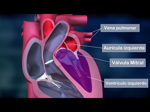
Anatomy of the Heart
- The heart consists of four chambers (cavities): two atria (upper chambers) and two ventricles (lower chambers).
- Atria serve as reservoirs for blood before passing it to ventricles; they have thin walls that withstand low pressure.
- Ventricles are muscular chambers that pump blood out of the heart; they contract forcefully due to numerous muscle fibers.
Separation and Coordination
- A fibrous wall called septum separates right and left sides of the heart, preventing blood mixing.
- Interatrial septum separates atria; interventricular septum separates ventricles.
- Atria fill with blood almost simultaneously before contracting to send it into ventricles.
Role of Valves in Blood Flow
- Four valves within the heart ensure unidirectional blood flow:
- They open to allow blood entry and close to prevent backflow.
- Two groups exist: semilunar valves (aortic and pulmonary valves), which manage flow from ventricles to arteries, and atrioventricular valves (mitral and tricuspid), which control flow between atria and ventricles.
Functionality During Cardiac Cycle
- Semilunar valves close during diastole when ventricles fill with blood.
- Aortic valve prevents backflow into left ventricle; pulmonary valve prevents backflow into right ventricle.
- Atrioventricular valves close during systole when ventricles contract, ensuring efficient pumping towards arteries.
Understanding the Circulatory System
Overview of Blood Vessels and Heart Function
- The veins delivering blood from the heart are the superior vena cava and inferior vena cava. The superior vena cava collects blood from the upper body, while the inferior vena cava gathers blood from below the diaphragm, both directing it to the right atrium.
- Two main arteries carry blood away from the heart: the pulmonary artery transports deoxygenated blood from the right ventricle to the lungs, and the aorta carries oxygenated blood from the left ventricle to all body tissues.
Circuits of Blood Flow
- Blood circulation occurs in two circuits:
- Pulmonary Circuit: Shortest circuit that moves deoxygenated blood to lungs for oxygenation.
- Systemic Circuit: Longest circuit transporting oxygenated blood throughout all body tissues.
Heart's Role in Pumping Blood
- The heart functions with atria as reception chambers and ventricles as expulsion chambers. Deoxygenated blood enters through superior/inferior vena cavas into right atrium, which contracts to send it to right ventricle via tricuspid valve.
- Upon contraction of right ventricle, blood is pushed into pulmonary artery towards lungs for oxygenation. This process requires opening of pulmonary valve for smooth passage before closing again.