Neurofisiología - Somatosensorial - UBA - MEDICINA
New Section
The section introduces the practical work on somatosensory perception, focusing on how the body gathers and processes information from both external and internal environments to generate specific responses.
Understanding Somatosensory Processing
- The nervous system processes tactile information, distinguishing between coarse touch and fine touch, with the latter referring to detailed sensations.
- Somatosensory processing includes proprioception (body part positioning), pain, and temperature perception, collectively known as thermal senses.
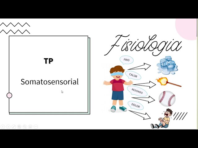
- Illustration of a child recognizing coldness, heat, shapes, and pain with eyes closed highlights how the body interprets sensory inputs.
Types of Stimuli in Somatosensory Perception
- Stimuli in somatosensory perception exhibit four main characteristics: modality, intensity, temporality, and spatiality.
- Modality refers to the type of stimulus received by receptors—physical, chemical, or other forms.
- Examples of stimuli include noxious (harmful), thermal (heat/cold), tactile (touch), and chemical stimuli like those causing pain.
Receptor Specialization and Transduction Process
- Receptors are specialized for different types of stimuli such as nociceptors for harmful stimuli or thermoreceptors for temperature changes.
- Receptors undergo transduction where they convert received stimuli into electrical energy for transmission to the central nervous system.
Intensity Coding in Somatosensation
- Intensity of a stimulus determines the sensation's strength; higher intensity leads to stronger bodily responses.
New Section
This section discusses the relationship between stimulus intensity and neuronal discharge frequency, highlighting how different stimuli impact neurotransmitter release.
Understanding Neuronal Response to Stimuli
- The frequency code indicates that higher intensity stimuli lead to increased neuronal discharge frequency.
- Stimulus intensity influences neurotransmitter release; gentle touch results in lower action potential frequency compared to a stronger stimulus like a punch.
Temporal Characteristics of Stimuli
This part delves into the temporal aspects of stimuli, exploring adaptation and habituation as crucial concepts in neural response dynamics.
Adaptation and Habituation in Neural Responses
- Temporal characteristics involve adaptation and habituation, affecting receptor responses.
- Adaptation involves reduced receptor response to constant stimuli, adjusting the response threshold at the peripheral nervous system level.
Adaptation Mechanisms in Neural Responses
This segment elaborates on adaptation mechanisms within neural responses, distinguishing between peripheral and central nervous system processes.
Differentiating Adaptation and Habituation
- Habituation occurs at the central nervous system level, reducing attention to specific stimuli over time for cognitive prioritization.
- Central habituation enables focusing on critical stimuli by desensitizing certain sensory inputs through cortical adjustments.
Receptor Response Dynamics
Exploring receptor response dynamics sheds light on adaptation types based on stimulus duration and intensity.
Receptor Types Based on Adaptation Speed
- Receptors exhibit fast or slow adaptation rates, impacting their responsiveness to dynamic or static stimuli.
Understanding Receptor Adaptation and Spatial Localization
In this section, the discussion revolves around receptor adaptation, distinguishing between receptors with slow adaptation and those with rapid adaptation. Additionally, the concept of spatial localization of stimuli is explored in relation to how the body recognizes the origin of a specific stimulus.
Receptor Adaptation
- Slow Adaptation Receptors:
- Constant discharge without adaptation.
- Continuous generation of action potentials upon stimulus application.
- Rapid Adaptation Receptors:
- Initially maximum discharge in response to a stimulus.
- Decreased discharge over time with persistent stimulation until it may disappear completely.
- Differences Between Slow and Rapid Adaptation:
- Slow adaptation gradually decreases action potential generation with continued stimulation.
- Rapid adaptation ceases action potential generation when the stimulus is removed but may sporadically generate some action potentials during stimulation.
Significance of Receptor Types
- Purpose of Different Adaptations:
- Slowly adapting receptors dedicated to pain and temperature perception for continuous vigilance over potentially harmful stimuli.
- Rapidly adapting receptors specialized for touch sensation to filter out non-critical stimuli for constant awareness without unnecessary attention.
Spatial Localization of Stimuli
- Understanding Spatial Recognition:
- Body's ability to identify the source of a specific stimulus.
- Example scenario illustrating how individuals discern touches on different body parts accurately.
- Mechanisms for Spatial Discrimination:
- Utilization of receptive fields for spatial coding.
- Topographical organization within the central nervous system aids in precise spatial discrimination.
Exploring Field Receptive Characteristics
This segment delves into field receptive properties, emphasizing their role in modulating receptor responses based on specific stimulations across various body regions.
Field Receptive Definition
- Defining Field Receptive Area:
- Region where stimulation enhances a single receptor's firing rate.
- Correspondence between tissue area and receptor activation for signal processing.
Variability in Field Receptive Sizes
- Size Variation Impact:
- Influence on firing frequency modulation based on stimulated region.
Understanding Receptive Fields in Sensory Systems
In this section, the speaker discusses the concept of receptive fields in sensory systems, focusing on how the size of these fields impacts spatial resolution and stimulus recognition.
Small vs. Large Receptive Fields
- : Small receptive fields have high specificity and spatial resolution, allowing precise identification of stimuli.
- : Large receptive fields lack specificity, leading to lower spatial resolution and difficulty in pinpointing stimulus location.
Determining Receptive Field Size
- : The threshold for two-point discrimination helps determine receptive field size.
- : Receptive fields are areas where stimulation increases receptor firing rates, crucial for spatial perception.
Impact on Spatial Resolution
- : Smaller receptive fields result in better spatial resolution due to lower two-point discrimination thresholds.
- : Larger receptive fields require greater distances between stimuli for separate recognition, affecting spatial perception.
Experimental Observations
- : Two-point stimulation experiments reveal differences in perceived stimuli based on receptive field sizes.
- : Varied responses to stimuli distances highlight how field size influences sensory processing.
Practical Implications and Understanding
- : Differences in tactile sensitivity between body regions showcase varying receptive field sizes' impact on stimulus recognition.
Understanding Receptive Fields and Sensory Processing
In this section, the speaker discusses the concept of receptive fields in sensory processing, emphasizing how different body parts have varying receptive field sizes and how stimulation affects these fields.
Receptive Fields and Stimulation
- Receptive fields vary in size across body parts.
- Different body parts have different receptive field sizes.
- Stimulation impacts receptor activation.
- Stimulating a single receptive field activates one receptor.
- Distances between stimuli affect receptor activation.
Spatial Resolution and Receptor Density
- Spatial resolution differs based on receptor density.
- Body regions have varying receptor densities and field sizes.
- Two-point threshold varies across body regions due to receptor characteristics.
- Two-point thresholds differ: tongue (2mm), shoulder (64mm), fingertips (4mm).
Organizational Strategies in Sensory Systems
This part delves into the organizational strategies within sensory systems, highlighting hierarchical processing, parallel processing, and somatotopy.
Hierarchical Processing
- Sensory systems employ hierarchical processing.
- Three types of organizational strategies exist: hierarchical/serial processing.
Parallel Processing
- Parallel processing is another organizational strategy for sensory information.
- Different modalities process simultaneously through separate pathways.
Somatotopy
- Somatotopy involves cortical representation of body areas based on sensitivity levels.
New Section
This section discusses the factors that determine spatial resolution in sensory perception, focusing on the density of receptors and receptive fields.
Factors Affecting Spatial Resolution
- The size of receptive fields influences spatial resolution; larger fields result in lower resolution, while smaller fields lead to higher resolution.
- Information travels through the nervous system differently based on receptor density and field size, affecting cortical representation.
- Body parts with high sensitivity, like hands and lips, have extensive cortical representation due to receptor density and field size.
- Receptor inheritance, field size, and cortical representation collectively determine spatial resolution in touch perception.
New Section
This part delves into somatosensory receptors responsible for collecting external and internal sensory information.
Somatosensory Receptors
- Somatosensory receptors gather information from both external and internal environments for transduction.
- Receptors specialize in converting various stimuli (touch, pain, temperature) into electrical signals for transmission.
- Receptors can be peripheral extensions of primary neurons or specialized cells based on the type of sensory input they receive.
New Section
This segment explores different types of somatosensory receptors associated with distinct modalities such as touch, proprioception, temperature, and pain.
Types of Somatosensory Receptors
- Each receptor corresponds to a receptive field; four main modalities are touch, proprioception, temperature, and pain.
- Mechanoreceptors like Merkel discs detect fine touch using Aβ fibers; proprioceptors monitor muscle position via α/β fibers.
Understanding the Sensory System
This section delves into the receptors of the somatosensory system, detailing their structure and function in processing sensory information.
Receptors of the Somatosensory System
- Sensory information from receptors is transformed into electrical energy for transmission.
- Sensory receptors are free nerve endings responsible for collecting and transmitting sensory data.
- The first neuron in the pathway processes sensory stimuli and transmits them centrally.
- Information travels through the dorsal root towards the central nervous system for recognition.
- Neurons pass through various relay stations to reach the cortex for stimulus processing.
Classification of Sensory Neurons
This segment categorizes sensory neurons based on axon characteristics, highlighting their role in different types of sensory perception.
Types of Sensory Neurons
- Primary sensory neurons can be classified based on axon characteristics such as touch, proprioception, temperature, and pain.
- Different fiber types (alpha, beta, delta, C fibers) vary in diameter and conduction speed.
- Alpha and beta fibers have high conduction speeds due to their large diameter.
- C fibers transmit information slowly due to their small diameter and myelination status.
Peripheral Nerves and Sensitivity
Discusses how peripheral nerves convey sensory information and potential implications of nerve damage on sensitivity.
Peripheral Nerves Functionality
- Severe peripheral nerve injuries can lead to complete loss of sensation in specific regions they innervate.
Neurological Pathways Overview
In this section, the discussion revolves around the primary afferent neurons that make up the peripheral nerve and their role in transmitting sensory information to the central nervous system. The concept of receptive fields, their non-overlapping nature, and how sensory information is processed through these pathways are explored.
Primary Afferent Neurons and Receptive Fields
- Primary afferent neurons constitute the peripheral nerve responsible for conveying sensory information to the central nervous system.
- Damage to a peripheral nerve results in loss of sensitivity in all receptive fields innervated by that nerve.
- Sensory information from different receptive fields travels through primary afferent neurons to plexuses where there is an intermingling of information before entering the spinal cord.
Information Processing in Neural Pathways
- Receptive fields do not overlap; each field receives input from a unique primary afferent neuron.
- Sensory information enters the spinal cord via dorsal roots, showing overlap from various receptive fields at this level.
Dermatomes and Sensory Loss
This segment delves into dermatomes, territories of skin innervated by dorsal roots of spinal nerves. The discussion highlights how lesions affecting these areas can lead to specific patterns of sensory loss on the skin.
Dermatomes and Sensory Alterations
- Dermatomes represent skin territories innervated by dorsal roots of spinal nerves post plexus crossover.
- Lesions on dorsal roots result in loss of sensation in corresponding dermatomal regions, aiding in identifying affected medullary sectors.
Clinical Implications and Sensory Mapping
This part focuses on clinical applications related to sensory loss patterns and their significance in diagnosing neurological conditions based on affected dermatomes or peripheral nerves.
Clinical Correlations
- Hematomas follow distinct transverse band patterns across body regions, aiding clinical assessment based on sensory deficits.
- Identification of sensory loss within specific dermatomes helps differentiate between central (spinal cord-related) and peripheral nerve lesions.
Specialization of Receptors
The discussion shifts towards receptor specialization for different modalities such as touch, pressure, proprioception, pain, and temperature. Each modality has distinct receptors specialized for its detection.
Receptor Specialization
- Receptors are specialized for particular modalities like touch, pressure, proprioception, pain, and temperature.
Detailed Overview of Mechanoreceptors in the Skin
In this section, the speaker discusses different types of mechanoreceptors found in the skin, their locations, structures, functions, and percentages in tactile receptors.
Mechanoreceptors Types and Functions
- Pazzini receptors are deep in the skin's dermis and have a unique structure with a capsule resembling onion layers. They sense deep pressure and vibrations rapidly.
- Merkel receptors are superficially located throughout the skin and hair follicles, especially abundant at fingertip endings. They play a key role in recognizing objects through touch and static pressure.
- Ruffini receptors are deep in the dermis, sensitive to skin deformation and stretching. They adapt slowly and constitute 20% of hand mechanoreceptors.
- Two mnemonic rules help differentiate receptor types: Germans (Meissner & Merkel) are superficial while Italians (Ruffini & Pazzini) are deep; Messi & Pele run fast indicating Meissner & Pazzini as rapid adapters.
Adaptation Characteristics and Importance
- Understanding receptor location aids in distinguishing their adaptation type. Sharing mnemonic rules can assist in memorizing these distinctions for effective learning.
- Differentiation between rapid (Meissner & Pazzini) and slow adapters (Merkel & Ruffini) is crucial for understanding sensory processing mechanisms.
Proprioception Receptors: Understanding Body Positioning
This part delves into proprioception receptors that provide detailed information on body mechanics, limb positions, movements' directionality, aiding precise movement control.
Proprioception Receptor Types
- Proprioceptors gather mechanical data about body functions like limb position awareness crucial for precise movement control.
- These receptors perceive body/members' positions (kinesthesia), movement directions/senses (synesthesia). Key proprioceptors include muscle spindles, Golgi tendon organs, joint receptors.
Muscle Spindle Functionality
- Muscle spindles detect muscle length changes indicating stretch levels during gross or fine movements. Muscles involved in finer actions like eye or hand muscles have higher spindle density.
Understanding Muscle Fiber Types and Neuromuscular Components
In this section, the speaker delves into the structure of muscle fibers, highlighting the differentiation between intrafusal fibers and extrafusal fibers. The discussion also covers the classification of muscle fibers based on nuclear arrangement and their relationship with neuromuscular components.
Muscle Fiber Structure and Classification
- Muscle fibers are surrounded by a capsule of connective tissue. Extrafusal muscle fibers form the striated muscle peripherally, while intrafusal muscle fibers are centrally located.
- Intrafusal fibers have a non-contractile central portion, with contractile ends termed as contractile ends. They can be classified based on nuclear arrangement into chain nuclear or bag nuclear types.
- The central region of intrafusal muscle fibers is closely related to primary afferent fibers that transmit information about muscular stretch to the central nervous system. Two types of afferent fibers, fiber 1a and type 2, play roles in conveying rapid responses to changes in muscle length.
Neuromuscular Components and Functions
- Motor neurons alpha innervate extrafusal muscle fibers for contraction, while motor neurons gamma innervate contractile ends of intrafusal fibers within the neuromuscular spindle.
- Motor neurons alpha generate muscular contractions by stimulating extrafusal muscle fibers directly. In contrast, motor neurons gamma control contractile ends of intrafusal fibers within the neuromuscular spindle.
Role of Golgi Tendon Organ and Joint Receptors in Muscular Function
This segment explores the functions of Golgi tendon organs located within tendons and joint receptors situated at articulations. These structures play crucial roles in sensing mechanical changes during movement.
Golgi Tendon Organ and Joint Receptors
- The Golgi tendon organ is positioned in series with extrafusal muscle fibers within tendons. It detects tension in muscles through type 1b afferent nerve endings characterized by high conduction velocity.
- Joint receptors sense mechanical deformations at joints before movements occur. They provide feedback on positional changes in body parts, contributing to joint mobility.
Temperature Receptors: Thermoreceptors
- Apart from touch and proprioception receptors, thermoreceptors respond to temperature variations. Cold receptors (Aδ-fibers) convey cooling sensations rapidly but have smaller diameters compared to warm receptors (C-fibers), which transmit heat sensations more slowly due to their small diameter.
Temperature and Nociceptors
In this section, the discussion revolves around temperature sensitivity and nociceptors, which are pain receptors responding to tissue damage caused by various stimuli.
Temperature Sensitivity
- Nociceptors respond to thermal, chemical, and mechanical stimuli induced by tissue damage.
- Termorreceptors are activated at temperatures above 45 degrees Celsius, leading to increased discharge frequency.
Nociceptors and Sensitization
- Nociceptors are free nerve endings branching along body tissues with two types of fibers: A-delta (small diameter, high conduction speed) and C fibers (small diameter, low conduction speed).
- Sensitization involves lowering the activation threshold of nociceptive receptors in response to damaging stimuli.
Mechanism of Sensitization
- Sensitization reduces the threshold for pain receptor activation, potentially causing spontaneous discharge even with minimal stimulation.
- The sensitization process is triggered by interactions between receptors and a group of chemicals known as "soup sensitizer" or inflammatory soup.
Inflammatory Soup Components
This section delves into the composition of the inflammatory soup released during tissue damage, comprising various substances aiding in protection and healing processes.
Composition of Inflammatory Soup
- Tissue damage leads to the release of immune cells' substances that promote tissue protection and healing.
- Vasodilation occurs in the damaged area to facilitate the influx of substances aiding in protection and scar formation.
Role of Inflammatory Soup Components
- Various components like neurotransmitters act on primary afferent nerve endings to sensitize them for easier recognition of stimuli.
New Section
In this section, the speaker discusses the role of various substances in sensitizing pain receptors and promoting pain sensation.
Understanding Pain Sensitization
- Histamine released by mast cells contributes to the sensitization process.
- Various substances, including histamine and substance P, play a role in sensitizing pain receptors.
- Damage to tissues leads to the release of these substances, affecting nociceptive nerve endings.
New Section
This part focuses on tissue damage and the substances involved in sensitizing pain receptors.
Tissue Damage and Sensitization
- The image illustrates tissue damage and the substances forming the sensitizing soup released due to cell rupture.
- Substances from various sources act on primary afferent nociceptors, influencing their sensitivity.
New Section
Exploring how substances interact with nociceptive terminals to sensitize them.
Interaction with Nociceptive Terminals
- Substances like histamine decrease activation thresholds of nociceptive terminals, facilitating easier activation.
- Sensitization at peripheral receptor sites contrasts with adaptation and habituation seen earlier in the video.
New Section
Contrasting sensitization with adaptation and habituation while explaining hyperalgesia.
Sensitization vs. Adaptation/Habituation
- Sensitization lowers activation thresholds at nociceptive terminals, increasing responses to painful stimuli.
- Hyperalgesia involves heightened sensitivity to pain due to decreased activation thresholds.
New Section
Classifying hyperalgesia into primary and secondary types based on location.
Classification of Hyperalgesia
- Primary hyperalgesia occurs at the site of injury due to inflammatory processes sensitizing local receptors.
- Secondary hyperalgesia can be peripheral (around the injury) or central (in the nervous system).
Perception of Pain and Sensitization
In this section, the discussion revolves around the sensitization process related to pain perception and its physiological mechanisms.
Peripheral Sensitization
- Peripheral sensitization lowers the activation threshold in response to stimuli.
- Substance P increases vasodilation and stimulates astrocytes to release histamine.
- These physiological processes aim to protect the injured area, promote healing, and defend against infections by increasing blood flow and immune cell migration.
Central Sensitization
- Central sensitization occurs in the second neuron of the pain pathway in the spinal cord.
- Hyperactivation of primary nociceptors leads to sensitization of second-order neurons, causing hyperalgesia.
- Hyperalgesia results in perceiving non-painful stimuli as painful (allodynia), indicating heightened sensitivity to pain signals.
Pain Perception Graph
- A graph illustrates pain intensity versus stimulus intensity.
- The dashed line represents the pain threshold; stimuli above it are perceived as painful, while those below are non-noxious.
- Hyperalgesia increases sensitivity to painful stimuli, while allodynia causes pain from typically non-painful stimuli.
Mechanisms of Hypersensitivity
This section delves into hypersensitivity mechanisms affecting pain perception thresholds.
Hyperalgesia vs. Allodynia
- Hyperalgesia amplifies sensitivity to painful stimuli beyond normal thresholds.
- It results in perceiving mild stimuli as painful due to lowered pain thresholds.
- Allodynia induces a painful response from usually innocuous stimuli.
- Even minor stimuli trigger pain responses below normal thresholds due to heightened sensitivity.
Neural Pathways
- Primary sensory neurons transmit information from receptors through peripheral and central pathways.
- Information is relayed via dorsal root ganglia towards the central nervous system for processing and interpretation.
New Section
In this section, the discussion revolves around the transmission of sensory information related to touch and temperature through different systems in the body.
Transmission of Sensory Information
- The lateral spinothalamic tract is responsible for carrying information regarding crude touch reception and temperature.
- The posterior column-medial lemniscus pathway transmits detailed touch information, also known as discriminative touch or proprioception.
- The dorsal column-medial lemniscus system recognizes fine touch, discriminating edges, textures, sizes, and details of touch.
Neuronal Pathways for Sensory Perception
- Mechanoreceptors and proprioceptive sensations are collected by the first-order neuron with a pseudo-unipolar structure.
- The first-order neuron's cell body is located in the dorsal root ganglion before transmitting sensory information centrally towards the spinal cord.
Organization of Sensory Pathways
- Somatotopic organization exists in the posterior columns where lower limb information ascends medially while upper limb, trunk, and neck data ascend laterally.
- Second-order neuron bodies maintain ipsilateral positioning in the cuneate and gracile nuclei at the bulb level, receiving sensory input from corresponding body regions.
Projection to Cortical Areas
- Axons from second-order neurons cross midline forming medial lemniscus before reaching thalamic nuclei contralaterally for further projection to somatosensory cortex.
- Sensory signals become conscious in the somatosensory cortex (Brodmann area 3) within the parietal lobe, representing tactile and proprioceptive stimuli from contralateral body areas.
New Section
In this section, the speaker discusses the importance of understanding the sensory system and its components.
Understanding the Sensory System
- The essential information for the upcoming exam includes details about the type of information received by the sensory system, characteristics of receptors, neuron locations in pathways, and specific sites crucial for processing.
- Knowledge about the bulbar region is vital for clinical applications, emphasizing its significance in medical practice.
- Collaterals from the first neuron's central extension at the spinal cord transmit proprioceptive information from both upper and lower limbs to form the spinocerebellar system.
- The spinocerebellar system comprises anterior and posterior spinocerebellar tracts responsible for conveying unconscious proprioception information to the cerebellum.
- Primary afferents forming these tracts leave collaterals in the posterior gray horn of the spinal cord, interacting with second neurons crucial for spinocerebellar functions.
New Section
This section delves into how proprioceptive information from upper and lower limbs is processed through different pathways towards the cerebellum.
Processing Proprioceptive Information
- Collaterals carrying proprioceptive data synapse with second neurons based on entry points at different spinal levels, distinguishing between upper and lower limb inputs.
- The axon of these second neurons traverses through specific spinal regions like anterior white commissure before forming distinct tracts towards superior cerebellar peduncles for further transmission to cerebral cortex.
- Detailed pathways involving various nuclei like cuneiform nucleus are highlighted as critical points where synaptic connections occur to ensure accurate transmission of proprioceptive signals.
New Section
This part emphasizes summarizing key concepts related to sensory systems discussed earlier for better retention and understanding.
Recapitulation of Sensory System Details
- A concise summary is provided to aid in revisiting essential topics covered previously regarding sensory pathways and their functional significance.
- Differentiation between ascending pathways handling pain, temperature sensitivity (thermoreception), coarse touch (proto-patic sensation), and their respective receptors is outlined for comprehensive knowledge acquisition.
New Section
In this section, the speaker discusses the representation of different neurons and their pathways in the spinal cord.
Representation of Neurons in the Spinal Cord
- The second neuron makes synapses with the body of the second neuron located in the posterior horn of the spinal cord.
- Watson descends to the level of the spinal cord through the anterior white commissure before ascending contralaterally to locate the body of the third neuron.
- Three main tracts are formed by interactions between neurons: spinothalamic tract, anterior spinothalamic tract, and lateral spinothalamic tract.
- The spinothalamic tract conveys information about crude touch and pressure, while the anterior spinothalamic tract carries information about fine touch.
- The lateral spinothalamic tract transmits information related to pain and temperature sensation.
Understanding the Ascending Pathways in the Spinal Cord
In this section, the speaker discusses the ascending pathways in the spinal cord, focusing on different systems specialized for various sensory functions.
Ascending Pathways Overview
- The spinocerebellar pathway transmits proprioceptive information from the lower limbs to the cerebellum. It is associated with unconscious deep sensation and is not related to behavioral or emotional changes.
- The spinothalamic tract carries discriminative and tactile aspects of pain sensation but does not connect to areas of the limbic system associated with emotions.
- The paleospinothalamic tract is responsible for emotional and affective aspects of pain perception and behavior responses to pain stimuli.
Understanding Pathway Localization
- Ascending pathways traverse different regions of the spinal cord, such as the lateral spinothalamic tract (blue) and medial lemniscal system (pink), each following distinct routes within the spinal cord.
- Lesions at specific levels of the spinal cord lead to sensory deficits on corresponding sides of the body due to differences in pathway localization and crossing over.
Implications of Spinal Cord Lesions
- A lesion affecting the dorsal column-medial lemniscal system results in loss of fine touch sensation on one side, while a lesion impacting the anterolateral system leads to diminished crude touch perception on that same side.
- Alterations caused by spinal cord lesions affect both sides of the body due to ipsilateral ascending pathways being affected, contrasting with supraspinal lesions where deficits are contralateral since all pathways have crossed before reaching higher brain centers.
Trigeminal Pathway: Sensory Information from Face
This part delves into how sensory information from facial regions is transmitted through distinct trigeminal pathways.
Trigeminal Pathway Details
- Sensory information from facial regions travels via separate trigeminal pathways compared to general somatosensory input from other body parts.
- The trigeminal nerve consists of three main branches – ophthalmic, maxillary, and mandibular – each innervating specific territories on the face.
Detailed Overview of Neurological Pathways
In this section, the speaker delves into the intricate details of neurological pathways, focusing on the locations and functions of different nuclei within the sensory system.
The Second Neuron in Sensory Pathways
- The body of the second neuron in the pathway can be located in any of the three sensory nuclei in the fifth cranial nerve.
- Nuclei involved include the mesencephalic nucleus for proprioceptive information and the spinal nucleus or trigeminal spinal nucleus for touch, pain, and temperature data.
Projection to Thalamic Nuclei
- Axons from these nuclei project to the contralateral posterior medial thalamic nucleus for further relay to somatosensory cortex.
- Emphasize understanding the type of information carried by these pathways, primarily related to facial sensations and their projection to specific thalamic nuclei.
Mechanisms of Inhibition in Neurology
This segment explores two key mechanisms of inhibition within neurological processes: distal inhibition occurring centrally and lateral inhibition at peripheral levels.
Distal Inhibition Mechanism
- Distal inhibition is a central mechanism involving descending control from higher brain centers to inhibit primary afferent neurons in the spinal cord gray matter.
- It serves to prioritize important stimuli over less critical ones, exemplified by endogenous analgesia as an instance of this inhibitory process.
Lateral Inhibition Mechanism
- Lateral inhibition operates peripherally, enhancing contrast between activated and less active neurons for precise stimulus localization.
- By inhibiting secondary neurons with lower stimulation levels, lateral inhibition sharpens focus on primary stimuli location through increased activation intensity.
Inhibition and Analgesia Mechanisms in the Central Nervous System
The speaker discusses how distal inhibition allows the central nervous system to selectively control the perception of stimuli, particularly focusing on pain modulation and analgesia mechanisms.
Distal Inhibition and Pain Perception
- Distal inhibition is primarily activated by intense pain stimuli, leading to analgesia.
- Soldiers in war scenarios exemplify this phenomenon by continuing physical activities despite sustaining injuries due to the activation of endogenous analgesic systems.
- The endogenous analgesic system prioritizes pain relief during extreme situations like running with a gunshot wound, showcasing the body's adaptive response to significant pain.
Neural Pathways for Pain Modulation
- Pain signals ascend through the anterolateral system towards different regions of the central nervous system.
- The pathway involves primary sensory information entering through the spinal cord's posterior root and synapsing with secondary neurons that ascend laterally.
- Secondary neurons leave collateral branches at various levels along the spinal cord, influencing pain modulation processes.
Endogenous Analgesia Mechanisms
- Collaterals release glutamate in specific areas of the central nervous system, stimulating enkephalinergic interneurons that inhibit further pain transmission.
- Enkephalinergic interneurons suppress GABAergic interneurons responsible for inhibiting endogenous analgesia, thereby enhancing pain relief mechanisms.
Endogenous Opioid Release and Pain Relief
The discussion delves into how endogenous opioids are released within certain brain areas to induce analgesia and alleviate pain perception.
Opioid Release Mechanism
- Inhibiting GABAergic interneurons leads to the liberation of endogenous opioid systems, promoting analgesic effects within specific brain regions.
- Stimulation of mesencephalic areas triggers the release of endogenous opioid peptides like enkephalins, contributing to overall pain modulation processes.
Neurotransmitter Interactions for Pain Relief
- Descending pathways from higher brain centers activate noradrenergic and serotonergic interneurons in lower spinal levels, further modulating pain perception.
New Section
In this section, the speaker discusses the mechanism of pain perception and modulation in the nervous system.
Mechanism of Pain Perception and Modulation
- The painful stimulus triggers a mechanism that ascends to higher levels in the nervous system before descending back down to inhibit the same painful stimulus.
- Endogenous opioid peptides, such as morphine, act on receptors to induce pharmacological analgesia mediated by external drugs.