🥇 MÉDULA ESPINAL 1/4 - (Configuración Externa e Interna) -Anatomía. ¡Explicación fácil!
Introduction to Spinal Cord Anatomy
Overview of the Video Series
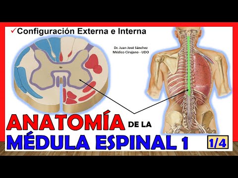
- This video is the first in a series of four on spinal cord anatomy, presented by Juan José Sánchez on Easy Anatomy.
- The focus will be on both internal and external configurations of the spinal cord. Subsequent videos will cover spinal roots, measurements, envelopes at the spinal level, and vascularization.
General Structure of the Nervous System
- The nervous system is divided into two main parts: the central nervous system (CNS) and peripheral nervous system (PNS). The CNS includes the brain (upper portion) and spinal cord (lower portion).
- The spinal cord is a continuation of the brain stem, consisting of thick bundles of nerve fibers. It is crucial to distinguish between spinal cord tissue and bone marrow; they serve different functions within the body.
Anatomical Protection and Surroundings
Meninges and Bone Structure
- The spinal cord is protected by membranes known as meninges, which are similar to those surrounding the brain. These membranes provide essential protection for the nervous tissue within.
- Additionally, there is a bony structure formed by vertebrae that offers mechanical protection to the spinal cord from external forces. Understanding this protective mechanism is vital for studying its anatomy effectively.
Curvatures and Limits of Spinal Cord
Curvature Characteristics
- The spinal cord exhibits several curvatures that correspond with those found in the vertebral column: cervical concavity posteriorly, thoracic convexity anteriorly, and lumbar concavity posteriorly. Understanding these curvatures aids in comprehending overall spine function.
Defining Limits
- The upper limit of the spinal cord connects with medulla oblongata; however, defining this boundary can be anatomically complex due to variations among individuals. It’s generally considered above where C1 emerges from this region.
Anatomy of the Spinal Cord
Overview of Spinal Cord Limits
- The upper limit of the spinal cord is defined by an imaginary line over the C1 vertebra (atlas) and the odontoid process of C2, separating the medulla oblongata from the spinal cord.
- The lower limit is identified at the intervertebral disc between L1 and L2, where the spinal cord narrows into a structure called conus medullaris.
Filum Terminale and Developmental Changes
- The filum terminale extends from the conus medullaris to either Co1 or Co2 vertebra, marking its endpoint in relation to coccygeal structures.
- In fetal development, at eight weeks, the spinal cord fills most of the vertebral canal; by 24 weeks, it rises to near S1; in newborns, it reaches L3; and in adults, it ends between L1 and L2 due to growth of the vertebral column.
External Morphology of Spinal Cord
- The spinal cord has a cylindrical shape that is slightly flattened anteroposteriorly but wider laterally. Its thickness varies along its length.
- It features two significant bulges: cervical intumescence (upper portion) and lumbar intumescence (lower portion), which are critical for nerve root formation.
Segmentation and Nerve Roots
- The average length of the spinal cord is about 45 centimeters. It segments into various myelomeres corresponding to different regions.
- The cervical region spans from C1 anterior arch to approximately C3 anterior arch. This area gives rise to several important nerves including those forming part of the cervical plexus.
Cervical Intumescence Details
- Cervical intumescence extends from around C3 to D3 vertebrae. It generates key nerves such as C4 (last cervical plexus nerve).
- This region also produces roots for brachial plexus nerves ranging from C5 to T1 (D1), crucial for upper limb innervation.
Thoracic Portion Insights
Understanding the Spinal Cord Anatomy
Overview of the Lumbosacral Intumescence
- The lumbosacral intumescence is a bulge in the spinal cord extending from approximately D9/D10 to L2 vertebrae, crucial for forming roots that contribute to both lumbar and sacral plexuses.
Conus Medullaris and Its Significance
- The conus medullaris represents the terminal part of the lumbosacral intumescence, located anteriorly at L2. It gives rise to sacral and coccygeal nerve roots.
Cauda Equina Formation
- Surrounding the conus medullaris is a collection of nerves known as cauda equina (horse's tail), which plays an essential role in innervating lower body structures.
Filum Terminale Structure
- The filum terminale extends from the tip of the conus medullaris downwards, featuring two portions: one within the dural sac (pia mater wrapped) and another outside it (dura mater enveloped).
External Anatomy of Spinal Cord
- The external anatomy includes three main faces: anterior, posterior, and lateral. Notably, the anterior face has a median fissure while posterior features grooves like posterior median sulcus.
Anterior Median Fissure Details
- The anterior median fissure runs along the midline; adjacent to it are anterolateral sulci where anterior roots of spinal nerves emerge. This area also contains pyramids leading to pyramidal decussation.
Posterior Surface Features
- On examining the posterior surface, there’s a posterior median groove corresponding with an anterior structure. Additionally, posterolateral sulcus marks where posterior roots emerge.
Fasciculus Separation in Spinal Cord
- The posterior intermediate sulcus divides white matter into two fascicles: gracile fasciculus (medial) and cuneiform fasciculus (lateral), critical for sensory pathways.
Lateral Face Characteristics
Internal Configuration of the Spinal Cord
Overview of Gray and White Matter
- The internal configuration of the spinal cord contrasts with that of the brain, where gray matter is peripheral and white matter is central. In the spinal cord, gray matter is central while white matter is peripheral.
Structure of Myelomers
- The spinal cord consists of segments known as metameric segments or myelomers, which give rise to spinal roots or nerves. Each segment contributes to a specific arrangement in gray matter.
Arrangement of Gray Matter
- The gray matter has an H-shaped structure characterized by lateral concavities. This shape allows for alignment across different spinal segments, forming three main columns: posterior, anterior, and intermediate columns around the central duct.
Anterior and Posterior Horns
- The anterior horn is more voluminous than the posterior horn and directs fibers obliquely towards white matter to form anterior roots of each spinal nerve. Conversely, posterior horns are thinner with a more regular contour, directing fibers laterally towards posterior roots.
Intermediate Column and Lateral Horn
- The intermediate column exists only at thoracic levels and contributes to the formation of the lateral horn, which contains nuclei related to the autonomic system—specifically sympathetic information from thoracic regions.
Commissures in Spinal Cord Anatomy
Gray Commissures
- Anterior horns are connected by an anterior gray commissure, located in front of the central canal; this structure plays a crucial role in integrating signals between both sides of the spinal cord. A similar structure exists for posterior horns called posterior gray commissure behind the central canal.
White Matter Composition
- Surrounding gray matter, white matter thickness varies along different sections; it is thicker at cervical levels but decreases as it descends through the spine. This variation affects how signals are transmitted throughout different regions of the body.
Cords within White Matter
Anatomy of the Spinal Cord
Structure of the Spinal Cord
- The posterior cord contains both the gracilis fasciculus medially and the cuneate fasciculus laterally, indicating their relative positions within the spinal cord.
- The lateral cord is located in the anterolateral sulcus and posterolateral sulcus, where posterior roots exit from the spinal cord.
Central Duct of the Spinal Cord
- The central duct, also known as the appendymal duct, runs along the entire length of the spinal cord and is covered by ependymal membrane.
- It serves as a continuation from the cerebral ventricles, beginning at the fourth ventricle and extending down to its termination point.