🥇 ANATOMÍA DE LA GLÁNDULA TIROIDES Y PARATIROIDES. Fácil, Rápida y Sencilla
Anatomy of the Thyroid and Parathyroid Glands
Introduction to the Video
- The video focuses on the anatomy of the thyroid gland and parathyroid glands, dedicated to Andrés Navarro and his family in Venezuela.
Anatomy of the Thyroid Gland
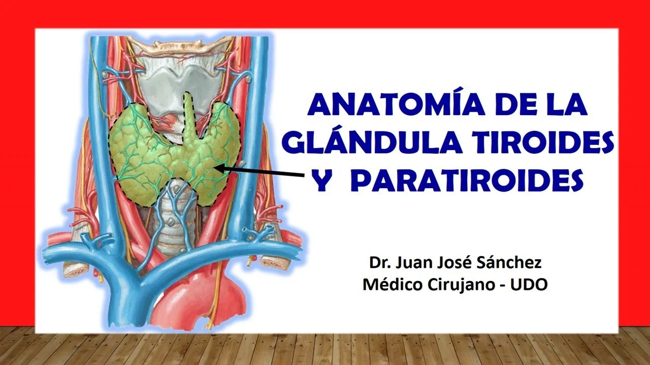
- The thyroid gland is named for its shield-like shape, derived from Greek etymology. It is located centrally in the neck, resembling an "H" or butterfly wings.
- Anatomically positioned between C5 and C7 vertebrae, with some sources suggesting it may extend to D1. The cricoid cartilage at C6 helps locate it.
- The thyroid gland has two protective capsules: an inner fibrous capsule attached directly to the gland and an outer false capsule from the deep cervical aponeurosis (pretracheal lamina).
- Comprised of two lobes connected by a central isthmus covering trachea rings 2, 3, and 4; may also have a pyramidal lobe that varies among individuals.
Structural Features of Each Lobe
- Each lobe has a vertex (top) directed backward, a base (bottom), and three faces: external, internal (medial), and posterior.
- The external face is covered by infrahyoid muscles like sternothyroid and omohyoid; this surface can be observed after sectioning sternocleidomastoid muscle.
Understanding the Thyroid Gland and Its Innervation
Anatomy and Blood Supply of the Thyroid Gland
- The superior laryngeal nerve, particularly its external branch, innervates the thyroid gland. The gland receives blood supply from two main arteries: the superior thyroid artery (a branch of the external carotid artery) and the inferior thyroid artery (from the bicervical scapular trunk).
- The superior thyroid artery has five branches, with glandular branches being most significant. These branches anastomose at the upper edge of the isthmus of the thyroid gland.
- A third, less common vessel known as the middle thyroid artery may also exist; it can originate from either the brachiocephalic trunk or right common carotid artery but is inconsistent in presence.
Venous Drainage and Lymphatic System
- Venous drainage involves three major veins:
- Superior thyroid vein (drains into internal jugular vein)
- Middle thyroid vein (also drains into internal jugular vein)
- Inferior thyroid vein (drains into brachiocephalic veins).
- Lymphatic drainage is categorized by anatomical lobes:
- Lobes drain upward to deep cervical nodes along internal jugular vein.
- Downward drainage occurs to paratracheal ganglia alongside trachea.
- The isthmus drains differently: upward towards pre-laryngeal ganglia in front of larynx and downward towards pretracheal ganglia in front of trachea.
Innervation and Hormonal Function
- The thyroid gland's innervation comes from both sympathetic and parasympathetic systems rather than cervical plexus due to its endocrine function. It produces hormones like T3, T4, and calcitonin.
- Sympathetic innervation arises from cervical sympathetic chain via superior cervical ganglion (along superior thyroid artery) and middle cervical ganglion (along inferior thyroid artery).
- Vagus nerve contributes to sympathetic innervation through recurrent laryngeal nerve and external branch of superior laryngeal nerve.
Surgical Considerations During Thyroidectomy
- In performing a total removal of the thyroid gland (thyroidectomy), surgeons must be cautious about three critical structures:
- Parathyroid glands which are small yet vital for life.
- Recurrent laryngeal nerve that controls most larynx muscles; injury can lead to hoarseness or speech issues.
Understanding the Parathyroid Glands
Overview of Parathyroid Glands
- The parathyroid glands are distinct from the thyroid gland, despite both being endocrine glands that secrete hormones into the bloodstream.
- They produce parathyroid hormone (PTH), which is essential for regulating calcium levels in the body and is crucial for life.
- There is some debate among medical literature regarding blood supply; while some sources state they are supplied solely by the inferior thyroid artery, others suggest that superior parathyroid glands may also receive blood from the superior thyroid artery.
Surgical Considerations
- During thyroidectomy procedures, it is vital to avoid damaging or removing the parathyroid glands, as they can be mistaken for lymph nodes.
- Awareness of their location and function is critical for surgeons to prevent complications related to calcium regulation post-surgery.
Additional Resources
- The speaker encourages viewers to explore more videos on related topics such as neck irrigation through various arteries, enhancing understanding of anatomical relationships.