🥇 ANATOMÍA DE LA LENGUA. ¡Explicación Fácil y Sencilla!
Anatomy of the Tongue
Introduction to the Tongue
- The video introduces the anatomy of the tongue, following previous discussions on oral cavity anatomy.
- The speaker celebrates reaching 100,000 YouTube subscribers and emphasizes the tongue's significance as a muscular organ composed entirely of striated muscle.
Muscular Structure and Function
- The tongue is anchored by powerful muscles that connect to the hyoid bone and lower jaw, preventing it from moving backward during swallowing.
- Additional muscular folds secure the tongue to various structures, including the temporal bone and pharynx.
Functions of the Tongue
- The primary functions of the tongue include:
- Swallowing: Facilitates pushing food bolus towards the pharynx.
- Taste: Houses taste buds for flavor perception.
- Chewing: Aids in mixing food against the palate during mastication.
- Speech: Essential for articulating words through its movements.
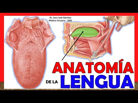
Overview of Tongue Anatomy
- The speaker encourages viewers to subscribe for more content and mentions that this video was highly requested.
- An overview of different parts of the tongue is introduced, including:
- Tip/Vertex: Front part extending towards incisor teeth.
- Back: Largest region viewed from above towards the palate.
Detailed Anatomical Regions
- Discussion on anatomical regions includes:
- Root: Connects to floor of mouth; serves as entry point for vascular and nervous structures.
Anatomy of the Tongue: Understanding Its Structure and Function
Overview of Tongue Portions
- The tongue is divided into two main portions: the buccal portion (anterior two-thirds) and the pharyngeal portion (posterior part, also known as the base).
- The buccal portion remains inside the mouth, while the pharyngeal portion is located in the oral pharynx. This distinction is often confused in anatomical texts.
Delimitation of Tongue Portions
- The cut-off point between these portions is marked by a V-shaped groove called the terminal sulcus. From this sulcus forward is the buccal portion; backward lies the pharyngeal portion.
- At both sides of the terminal sulcus, there exists a blind foramen known as foramen cecum, which represents remnants of embryological structures related to tongue development.
Lingual Papillae Types
- The dorsal region of the tongue features various types of lingual papillae, crucial for taste sensation:
- Filiform Papillae: Most abundant, pointed conical shape with tips facing upwards. They are primarily responsible for texture perception rather than taste.
- Fungiform Papillae: Located towards the tip and lateral edges; they have a mushroom-like appearance with a narrow base and wider top.
- Circumvallate Papillae: Ranging from 3 to 14 in number, these are found just anterior to the terminal groove and are characterized by surrounding depressions that house glands secreting saliva.
- Foliate Papillae: Found at the posterior part of the tongue; they appear as wrinkles and contribute minimally to taste but may have some lymphoid function.
Pharyngeal Portion Features
- The pharyngeal portion contains structures resembling lingual papillae but serves a lymphoid function instead of gustatory purposes; these form part of Waldeyer's ring along with other tonsils in the neck area.
- Mucosal folds connect this area to other structures like epiglottis through glossoepiglottic folds, creating spaces known as epiglottic valleculae on either side.
Microscopic Structures and Functions
- A microscopic view reveals distinct types of papillae:
- Lingual tonsils contain lymphoid tissue.
- Filiform papillae dominate in abundance.
- Fungiform papillae exhibit their characteristic mushroom shape.
- Circumvallate papillae are surrounded by glandular depressions that secrete saliva onto taste buds for enhanced flavor detection.
Lower Surface Characteristics
Understanding the Anatomy of the Tongue
Overview of Tongue Structure
- The tongue features folds known as fringed folds, located at the junction of its lower surface and the floor of the mouth, where major salivary gland ducts are found.
- The two medial orifices beside the lingual frenulum are openings for Wharton ducts, with one duct leading from the maxillary gland and others from sublingual glands.
- The deep artery of the tongue, also called the ranine artery, is visible on the underside; it appears purple when viewed from beneath.
Glands and Muscles Associated with the Tongue
- Anterior lingual glands, which produce saliva, are located on the underside of the tongue. The root connects to a set of muscles that facilitate movement.
- The hyoid bone and submaxillary gland are important anatomical landmarks; intrinsic muscles make up most of the tongue's structure while extrinsic muscles provide greater mobility.
Intrinsic Muscles of the Tongue
- There are four groups of intrinsic muscles: upper longitudinal (odd), lower longitudinal, transverse fibers, and vertical fibers. These allow for various movements including rolling in different directions.
- A fibrous structure referred to as a clear lingual septum acts as a partial divider within the tongue but does not completely separate it into halves.
Extrinsic Muscles and Their Functions
- Five extrinsic muscles include genioglossus muscle originating from mandible; it helps depress and protrude (stick out) the tongue.
- Understanding how these muscles function is crucial for airway management in medical scenarios; protruding the tongue can help clear airways during emergencies.
Importance of Anatomical Terms
Anatomy and Function of Tongue Muscles
Hyoglossus Muscle
- The hyoglossus originates from the greater horn and body of the hyoid bone, extending upwards to the lateral edge and lower surface of the tongue. Its contraction results in retraction of the tongue, opposing protrusion.
Styloglossus Muscle
- This muscle arises from the styloid process of the temporal bone and maxillary style ligament, intermingling with other tongue muscles. It inserts into the lateral edge of the tongue, functioning similarly to hyoglossus by retracting the tongue when contracted.
Palatoglossus Muscle
- Originating at the soft palate's palatine aponeurosis, this muscle also inserts into the lateral edge of the tongue. It plays a role in closing both anterior pillars during oropharyngeal contraction.
Chondroglossus Muscle
- A less commonly referenced muscle that originates from a minor part of the hyoid bone, often difficult to locate in anatomical atlases. It is theorized to reach towards the back of the tongue but does not extend fully there. The name reflects its cartilage origin (chondrus).
Tongue Irrigation and Innervation
Innervation and Lymphatic Drainage of the Tongue
Sensory and Gustatory Innervation
- The glossopharyngeal nerve (ninth cranial nerve) provides sensory and gustatory innervation to the posterior third of the tongue, along with contributions from the facial nerve through its lingual branch.
- The anterior two-thirds of the tongue receive sensory innervation primarily from the trigeminal nerve via the lingual nerve, while taste sensation is also facilitated by branches from the facial nerve.
- The vagus nerve contributes to sensory innervation in the posterior third through its internal branch of the superior laryngeal nerve, alongside fibers from both glossopharyngeal and facial nerves.
- It is noted that while discussing innervation, not all branches were illustrated; specifically, those from the facial nerve aiding gustation in the posterior third were omitted in visual representations.
Lymphatic Drainage
- Understanding lymphatic drainage is crucial as it directs flow to superficial cervical nodes such as submental and submaxillary nodes.