Fluorescent In Situ Hybridization (FISH) Assay
Introduction to Fluorescent In Situ Hybridization (FISH)
What is FISH?
- Fluorescent in situ hybridization (FISH) is a molecular cytogenetic technique that utilizes fluorescent probes to bind specifically to parts of chromosomes with high sequence complementarity.
- The DNA structure consists of two strands coiled into a double helix, where bases on each strand can bind together, allowing for hybridization between complementary sequences.
Mechanism of FISH
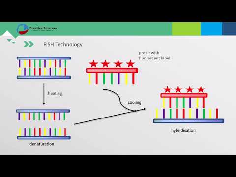
- FISH exploits the ability of one DNA strand to hybridize with another by using small DNA strands called probes that are labeled with fluorescent tags. These probes are complementary to specific chromosome regions.
- When DNA is heated, the strands denature and separate, enabling the probes to hybridize with their corresponding sequences in the patient's DNA. If there’s a deletion or duplication in the target region, it affects probe binding efficiency.
Types of Probes and Their Characteristics
- Different types of probes include double-stranded DNA (dsDNA), single-stranded DNA (ssDNA), and RNA probes:
- dsDNA probes are stable and easier to obtain.
- ssDNA probes offer higher specificity and resistance against degradation.
- RNA probes have better thermal stability and tissue penetration capabilities.
- Probes can be labeled either with radioactive isotopes or non-radioactive labels like biotin and fluorescent dyes, which influence detection methods used later in the process.
Steps Involved in FISH
- Preparation of Fluorescent Probes: Initial step involves creating suitable fluorescently labeled probes.
- Denaturation: The target DNA is denatured to allow probe binding.
- Hybridization: Probes hybridize with their complementary sequences within the target DNA.
- Detection: After hybridization, results are examined using fluorescence microscopy; preventing quenching during this step is crucial for accurate results.
Applications of FISH
- FISH has diverse applications across various fields including:
- Morphology
- Pathology
- Developmental biology
- Karyotyping
- Phylogenetic analysis