MENINGES E CIRCULAÇÃO LIQUÓRICA - PARTE 2
Overview of the Central Nervous System
Basic Structure and Components
- The speaker presents a simple representation of the central nervous system, highlighting a horizontal plane (in blue) that intersects with the foramen magnum of the occipital bone.
- Structures above this horizontal plane are located within the skull and constitute the brain, while the caudal continuation is found in the vertebral canal, protected by vertebrae.
Telencephalon and Diencephalon
- The telencephalon is noted to grow significantly, eventually corresponding to the diencephalon.
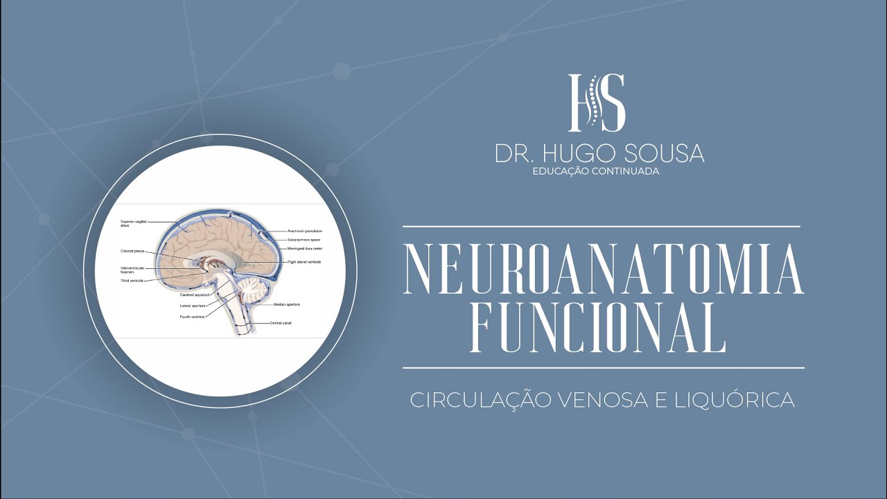
- Ventricular cavities within the telencephalon include lateral ventricles on both sides and a third ventricle at the center of the diencephalon.
Communication Between Ventricles
- There is communication between lateral ventricles via an interventricular foramen (Monro), leading to a third ventricle.
- The connection from the third ventricle to the fourth ventricle occurs through a mesencephalic aqueduct (Sylvius).
Ventricular System and Cerebrospinal Fluid
Fourth Ventricle and Spinal Canal
- The fourth ventricle is positioned posteriorly to the pons and anteriorly to the cerebellum, continuing into the central canal of the spinal cord.
Ependymal Cells and CSF Production
- Ependymal cells line ventricular cavities; they produce cerebrospinal fluid (CSF), also referred to as liquor or LCR.
Meninges Associated with CNS
Meningeal Layers
- The pia mater adheres closely to all parts of the central nervous system; it continues as a terminal filament that anchors in connective tissue at sacral hiatus.
Arachnoid Mater Representation
- Arachnoid mater surrounds CNS structures with trabeculae connecting it to pia mater throughout its extent.
Dura Mater Structure
Dual Layering of Dura Mater
- Dura mater has two layers: an outer periosteal layer associated with cranial bones and an inner meningeal layer that forms sinuses like superior sagittal sinus.
Sinuses Formation
- These layers create triangular-shaped sinuses lined by endothelium; they play crucial roles in venous drainage from brain structures.
Protection Mechanisms for CNS
Cranial Protection
- The cranium protects encased brain structures while vertebrae safeguard spinal cord components.
Periosteum Interaction
Understanding the Spaces Surrounding the Spinal Cord
Overview of Meningeal Spaces
- The periosteum and dura mater create a close relationship, leaving no space between them in certain areas, particularly around the spinal cord.
- The extradural space (also known as epidural or peridural space) is located around the spinal cord, providing a crucial area for various medical procedures.
- Unlike the brain, there is an extradural space present between bone and dura mater in the spinal region but not in cranial regions.
Subdural and Subarachnoid Spaces
- A subdural space exists between the dura mater and arachnoid layer surrounding the spinal cord; this space is more pronounced in cranial meninges.
- The subarachnoid space lies between arachnoid and pia mater layers, containing trabeculae that support its structure. This area is significant for cerebrospinal fluid (CSF).
Cerebrospinal Fluid Dynamics
- The subarachnoid space contains a large volume of cerebrospinal fluid (CSF), essential for cushioning and protecting the central nervous system.
- While there is minimal CSF in the subdural space, it serves different functions compared to other spaces like extradural which contains adipose tissue and venous plexus.
Clinical Relevance of Extradural Space
- The extradural/peridural/epidural spaces are clinically relevant due to their association with anesthetic procedures commonly referred to as "peridurals."
Choroid Plexus Functionality
- Choroid plexuses invade various ventricular structures within the brain, playing a critical role in producing CSF.
- Approximately 450 ml of CSF is produced daily by choroidal plexuses associated with ependymal cells across different ventricles.
Pathway of Cerebrospinal Fluid Flow
- CSF flows from lateral ventricles through interventricular foramina into third ventricle where it combines with additional production from third ventricle's choroidal plexus.
- After passing through cerebral aqueduct into fourth ventricle, CSF exits via median aperture or lateral apertures into subarachnoid spaces.
Understanding the Central Nervous System and Its Protection
The Role of Cerebrospinal Fluid (CSF)
- The cerebrospinal fluid (CSF) occupies the subarachnoid space surrounding the central nervous system, providing essential protection.
- CSF acts as a cushion against mechanical shocks, adhering to Pascal's principle, which states that pressure applied to a fluid is transmitted equally throughout.
- According to Archimedes' principle, any body immersed in a fluid experiences an upward buoyant force, making it effectively lighter and reducing trauma from contact with surfaces.
Production and Absorption of CSF
- CSF is produced within the central nervous system at a rate of approximately 450 ml per day in adults; around 125 to 150 ml is present in the neuroaxis at any time.
- The absorption of CSF occurs through arachnoid granulations projecting into the superior sagittal sinus, allowing for drainage into venous systems.
Venous Drainage Pathways
- The superior sagittal sinus connects laterally with transverse sinuses and sigmoidal sinuses, ultimately draining into internal jugular veins.
- Dural sinuses serve dual purposes: they facilitate both cerebrospinal fluid drainage and venous blood drainage from the central nervous system.
Anatomical Structures Involved
- A simple representation shows how arachnoid granulations project into the superior sagittal sinus for effective drainage.
- The internal structure includes layers of dura mater forming sinuses; these structures are crucial for maintaining proper cerebral function by managing both blood and CSF flow.
Clinical Implications
Neuroanatomy Insights
Injection of Polymerizable Resin
- The process involves injecting polymerizable resin to fill spaces, specifically passing through the interventricular foramen.
- If filling is unnecessary, infiltration of resin on one side is performed to ensure all areas are filled.
Examination of Lateral Ventricles
- After preparation, the brain is placed in an acidic medium to digest organic matter and obtain a mold.
- Observations include the large lateral ventricle associated with the telencephalon, highlighting its anterior portion (body), posterior horn, and inferior projection.
Third Ventricle Overview
- The third ventricle is centrally located within the diencephalon, featuring thalamic regions and interthalamic adhesion.
- The hypothalamus region includes optic recesses and infundibular recesses; it also contains the epithalamus associated with the pineal gland.
Connections Between Ventricles
- The connection between the third ventricle and fourth ventricle occurs via the cerebral aqueduct.
- Notable structures include median openings in the roof of the fourth ventricle leading to lateral apertures (foramina of Luschka).
Choroid Plexus Projections
- Observations reveal projections from choroid plexus at lateral apertures in addition to other anatomical features like bulbar olives and pyramids.
- The basilar artery's vascularization is discussed alongside choroid plexus projections in relation to cerebrospinal fluid pathways.
Cerebellar Anatomy
- Description includes both cerebellar hemispheres and their posterior aspects; mentions median openings allowing cerebrospinal fluid flow into subarachnoid spaces.