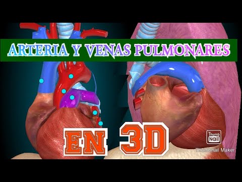
ANATOMIA 3D | ARTERIAS Y VENAS PULMONARES FÁCIL EXPLICACIÓN!
Anatomy of Pulmonary Arteries and Veins
Overview of Pulmonary Circulation
- The video discusses the anatomy of pulmonary arteries and veins, including their origins, relationships, terminations, and associated pathologies.
- Two types of blood supply to the lungs are highlighted:
- Nutritional irrigation from bronchial arteries (two for the left lung from the aorta; one for the right lung from a posterior intercostal artery).
- Functional irrigation responsible for gas exchange via pulmonary arteries and veins.
Structure and Function of Pulmonary Artery
- The pulmonary artery carries semi-deoxygenated blood (70% oxygen, 30% carbon dioxide) from the heart to the lungs.
- It serves as a conduit for blood expelled by the right ventricle towards the lungs.
- Originates from the right ventricle, forming a single trunk that divides into left and right pulmonary arteries.
Anatomical Features
- The pulmonary artery has valves at its origin known as Morgan's nodules; important for exam purposes.
- The trunk measures approximately 5 centimeters in length and is located between the second and third costal cartilages.
Pressure Dynamics
- Normal pressure in the pulmonary artery is around 16 mmHg; hypertension occurs when it exceeds 25 mmHg.
- A palpable pulse can be felt at this site due to arterial contraction.
Division into Left and Right Pulmonary Arteries
- After traveling about 5 cm, it bifurcates into left and right branches entering each lung's hilum.
- The structure entering each hilum includes:
- Pulmonary artery,
- Pulmonary veins,
- Main bronchus,
- Bronchial arteries,
- Bronchial veins,
- Pulmonary plexus nerves.
Hilum Anatomy Differences Between Lungs
- For the right lung:
- Entry order is main bronchus first (B), then pulmonary artery (A), followed by two pulmonary veins (V).
- For the left lung:
- Entry order is reversed: first enters with an artery (A), then main bronchus (B), followed by two pulmonary veins.
Bronchial Distribution and Pulmonary Segments
Overview of Bronchial Structure
- The bronchi divide into main bronchi, which further branch into lobar bronchi for the upper, middle, and lower lobes in the right lung.
- The right lung has two fissures (major and minor), dividing it into three lobes: superior, middle, and inferior.
Right Lung Segmental Anatomy
- The superior lobe contains three segments: apical, anterior, and posterior.
- The middle lobe consists of two segments: lateral and medial.
- The inferior lobe has five segments: apical, anterior, posterior, lateral, and medial.
Left Lung Structure
- The left lung has one fissure dividing it into two lobes: superior and inferior.
- The superior lobe features four segments: apical-posterior, anterior, lingular superior (related to cardiac notch), and lingular inferior.
Inferior Lobe Details
- In the left inferior lobe, only the anteromedial segment is visible from an anterior view; all other segments are observed laterally.
Pulmonary Arteries and Gas Exchange
Pulmonary Artery Pathway
- The pulmonary artery runs within the bronchial structure; it's described as intra-segmental due to its location relative to alveoli.
Gas Exchange Process
- Deoxygenated blood from the pulmonary artery enters alveoli for gas exchange with oxygen-rich air; this process results in oxygenated blood returning via pulmonary veins.
Pulmonary Veins Functionality
Structure of Pulmonary Veins
- There are three pulmonary veins on the right side (one for each lobe), while on the left side there are also two veins corresponding to each lobe.
Clinical Correlation with Heart Function
- Blood from pulmonary veins drains into the left atrium before moving to the left ventricle. Issues like left heart failure can lead to increased pressure in these veins.
Consequences of Left Heart Failure
Impact on Pulmonary Circulation
- Left ventricular dysfunction causes blood accumulation in the left atrium leading back to lungs through pulmonary veins. This can result in fluid leakage into alveoli causing acute pulmonary edema.
Pressure Measurements