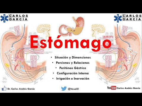
Anatomía - Estómago Part. I (Situación, Relaciones, Porciones, Peritóneo Gástrico)
Understanding the Stomach: Functions and Anatomy
Introduction to the Stomach
- The stomach is defined as a dilated portion of the digestive tube between the esophagus and duodenum. It serves three primary functions: storage, secretion, and gastric emptying.
Functions of the Stomach
- Storage: The stomach can hold 0.8 to 1.5 liters of ingested food, acting as a reservoir before digestion begins.
- Secretion: It produces gastric juice, including hydrochloric acid, which helps in breaking down food into chyme (a semi-liquid mass). This process transforms solid food into a form that can be absorbed in the intestines.
- Gastric Emptying: Once chyme is formed, it is gradually released into the small intestine for nutrient absorption.
Topographical Location of the Stomach
- The stomach occupies both the epigastric region and left hypochondrium within a cavity known as "space supracolic." Its boundaries include:
- Superiorly by diaphragm and liver.
- Inferiorly by transverse colon.
- Medially by celiac region.
Fixation Mechanisms of the Stomach
- The stomach's fixation involves its continuity with the esophagus at its upper end, where it adheres to the diaphragm through muscular fibers.
- Additionally, it has strong attachments at its posterior aspect near the greater curvature to maintain stability within abdominal cavity.
Dimensions and Variability of the Stomach
- Length: Approximately 25 cm from cardia to pylorus.
- Width: About 12 cm; thickness around 8 cm.
- Capacity varies significantly among individuals based on body type (e.g., thinner individuals have smaller stomachs compared to those who are obese).
Anatomical Parts of the Stomach
Vertical Portion
- Comprises several parts:
- Fundus: Dome-shaped structure fitting against diaphragm; contains air in radiographic images indicating normalcy.
- Forms an angle with esophagus known as "angle of His," crucial for preventing gastroesophageal reflux when angled correctly at about 45 degrees.
Body Structure
- The body resembles an inverted funnel; wider at top than bottom with significant directional change at angular incisure leading towards horizontal section.
Horizontal Portion
- Contains two segments:
- Antrum Pyloricum (pyloric vestibule).
Anatomy of the Stomach and Its Surroundings
Overview of the Pyloric Region
- The pyloric canal, or pylorus, is the widest part of the horizontal section of the stomach, serving as a pathway before entering the duodenum.
- The pyloric sphincter is a thickening of stomach muscles that acts as a valve for gastric emptying. This structure is crucial for regulating food passage into the small intestine.
Anatomical Boundaries
- A notable anatomical feature between the pylorus and duodenum is a marked depression known as the duodenal groove, which indicates where the stomach ends and the duodenum begins.
- The stomach has two surfaces: an anterior face and a posterior face, along with two curvatures: lesser curvature and greater curvature. Each plays a role in its anatomical relationships.
Anterior Surface Relations
- The anterior surface consists of both thoracic (in contact with ribcage) and abdominal portions; this division aids in studying its anatomy effectively.
- The thoracic portion lies behind ribs 5 to 9, relating closely to an important semiological space known as "semi lunar space," which has clinical significance in diagnostics.
Clinical Significance of Semi Lunar Space
- The upper limit follows the greater curvature from gastric fundus down to an imaginary line from xiphoid process to rib 9's apex along the left costal arch; this area can produce different sounds upon percussion based on organ density (hollow vs solid).
- In normal conditions, hollow organs like stomach produce tympanic sounds while solid organs like liver yield dull sounds during percussion examinations. Understanding these differences is vital for clinical assessments.
Abdominal Portion Relations
- The abdominal portion relates closely to anterior abdominal wall structures and liver; it forms a triangular area known as "triangle of Ave" defined by specific rib margins and an imaginary line across rib 9's apexes on both sides.
- Distinguishing between "triangle of Ave" and "semi lunar space" is essential for accurate anatomical understanding; each serves different diagnostic purposes in medical practice.
Posterior Surface Relations
- The posterior surface forms part of the anterior wall of omental bursa (lesser sac), covered mostly by peritoneum, indicating its relationship with surrounding organs such as diaphragm, adrenal glands, kidneys, pancreas, spleen, and transverse colon.
Anatomy of the Stomach and Its Surrounding Structures
Relationships of the Stomach
- The anterior face of the stomach is closely related to various structures, including the diaphragm, left adrenal gland, left kidney, pancreas, transverse colon, and spleen.
- The greater curvature of the stomach connects to several structures via peritoneal folds: it attaches to the diaphragm through the gastrofrénico ligament and to the spleen via the gastroesplénico omentum.
- Important vascular structures surround the greater curvature: arterial and venous arches as well as gastric lymph nodes are present around this area.
Lesser Curvature Connections
- The lesser curvature is connected to the liver by a fold known as the lesser omentum; this will be discussed in more detail later.
- Key vascular elements around this region include right and left gastric arteries along with their corresponding veins and lymphatic plexuses.
Peritoneal Coverage of the Stomach
- The stomach is almost entirely covered by peritoneum except for a small area at its fundus that adheres closely to the diaphragm—this area lacks peritoneal coverage.
- There are three main omenta associated with the stomach: greater omentum (gastrocolic), middle omentum (gastroesplénico), and lesser omentum (gastrohepático).
Description of Lesser Omentum
- The lesser omentum extends from the right border of abdominal esophagus, lesser curvature of stomach, and first part of duodenum down to a groove on visceral surface of liver.
- It consists mainly of two ligaments: hepatogastric ligament and hepatoduodenal ligament. This distinction is crucial as it often causes confusion in terminology.
Anatomical Features of Lesser Omentum
- The anatomical description shows that it has a quadrilateral shape with two surfaces (anterior/posterior) and four borders.
- The anterior surface is covered by visceral liver tissue; thus, visualizing it requires lifting up part of the liver.
Borders of Lesser Omentum
- Four borders are identified:
- Gastroduodenal Border: connects esophagus' right side with lesser curvature & duodenum's first portion.
Hepatic Pedicle and Greater Omentum Overview
Anatomy of the Hepatic Pedicle
- The liver has a relatively small diaphragmatic border, with a significant free edge known as the "borde libre" or pedicular border, which contains elements of the hepatic pedicle.
- The hepatic pedicle consists of three main components: the portal vein, the common bile duct (conducto colédoco), and the proper hepatic artery.
- The arrangement of these structures is crucial; the portal vein is located posteriorly, while the common bile duct is anterior-right and the proper hepatic artery is anterior-left.
Functionality and Structure
- The free edge contributes to forming the anterior lip of Winslow's recess (hiato de Winslow), which plays a role in abdominal cavity organization.
- The greater omentum, also referred to as "gastrocolic ligament," covers intestinal loops and serves as a protective layer during digestive surgeries.
Description of Greater Omentum
- This peritoneal fold originates at the greater curvature of the stomach, descending to approximately L5 vertebra before folding back up to attach to the transverse colon.
- It acts as a barrier that protects intestines by covering them and can help isolate infections within abdominal cavities.
Vascular Supply
- The gastro-splenic ligament connects to vessels supplying blood to both stomach and spleen; it includes splenic arteries traveling towards their respective organs.
Internal Configuration of Stomach
- The internal surface of a living stomach appears red but turns brownish-gray post-mortem due to rapid mucosal changes.
- Gastric folds (pliegues gástricos), more prominent in lower body regions than in fundus, contribute significantly to gastric surface area for digestion.
Cardiac Region Insights
- At cardia level, there exists an inconsistent mucosal fold known as "valvula cardiaca," which lacks anatomical sphincter function despite its name.