Clase 56 Fisiología Renal - Micción urinaria (IG:@doctor.paiva)
56th Class of Physiology: Urinary Mission
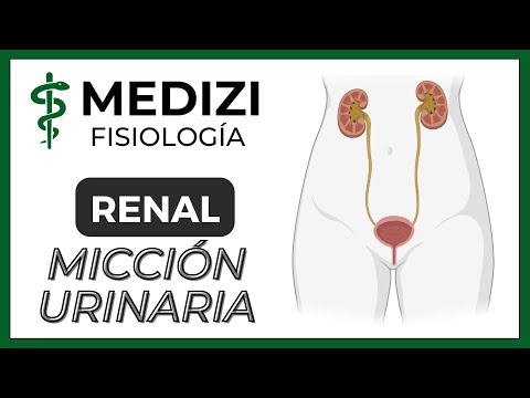
Overview of the Urinary System
- The urinary system consists of two kidneys, ureters, a urinary bladder, and the urethra. The process begins with nephron filtration, absorption, and secretion leading to urine formation.
- Urine exits through the renal papillae and travels via minor calyces to major calyces, then to the renal pelvis before reaching the ureters and being stored in the bladder.
Mechanism of Micturition
- Micturition is defined as the process by which the bladder empties when full. It occurs in two phases: filling until tension exceeds a threshold and triggering a reflex response known as micturition reflex.
- This reflex is primarily spinal (medullary) but can be influenced by higher brain centers that either inhibit or facilitate it.
Anatomy of the Bladder
- The bladder comprises smooth muscle (detrusor) responsible for urine storage and expulsion; it has two main parts: body (storage area) and neck (leading to urethra).
- Detrusor muscle contraction raises bladder pressure significantly (40-60 mmHg), essential for effective emptying during micturition. Its fibers are interconnected via gap junctions allowing synchronized contractions.
Triangular Area in Bladder
- A triangular region called trigone is formed where ureters enter at its upper angles; this area plays a crucial role in bladder function.
- The neck contains an internal sphincter (involuntary control) that maintains basal tone to prevent involuntary urination while storing urine. An external sphincter exists as skeletal muscle under voluntary control for conscious urination management.
Nervous System Control
- Innervation of the bladder comes from pelvic nerves connecting it to spinal segments S2-S3; these include sensory fibers detecting wall distension initiating micturition reflexes and motor fibers affecting detrusor contraction.
- Additionally, sympathetic innervation influences blood vessel stimulation rather than direct contraction/relaxation effects on detrusor muscles; autonomic nervous system regulates involuntary aspects of micturition including detrusor contraction and sphincter relaxation or constriction based on stimuli received.
Urine Composition During Storage
Understanding the Mechanisms of Urination
The Role of Stretching in Bladder Function
- Stretching increases pacemaker function and enhances peristaltic contractions in adults. The average length of ureters is approximately 25 to 35 centimeters, entering the bladder through the detrusor muscle.
Detrusor Muscle and Urine Flow Regulation
- The basal tone of the detrusor muscle prevents retrograde urine flow into the kidneys, maintaining a one-way system for urine expulsion. This is crucial for proper urinary function.
Micturition Reflex Activation
- Upon bladder distension, signals are sent to the spinal cord via pelvic nerves, triggering parasympathetic responses that cause detrusor contraction. These contractions typically last about a minute when bladder volume is low but increase as filling occurs.
Auto-regenerative Nature of Micturition Reflex
- As bladder volume increases, initial contractions activate sensory receptors leading to heightened reflexive impulses that further enhance detrusor contractions—a self-amplifying cycle until relaxation occurs after significant pressure build-up.
Inhibition of External Sphincter During Micturition
- A strong micturition reflex can inhibit voluntary control over the external sphincter; if this inhibition surpasses voluntary constriction efforts due to high bladder distension, urination occurs involuntarily. Signals also reach higher brain centers which can modulate this reflex based on situational appropriateness.
Voluntary Control Over Micturition