Western Blot Method - Animated Video
Western Blotting Technique Overview
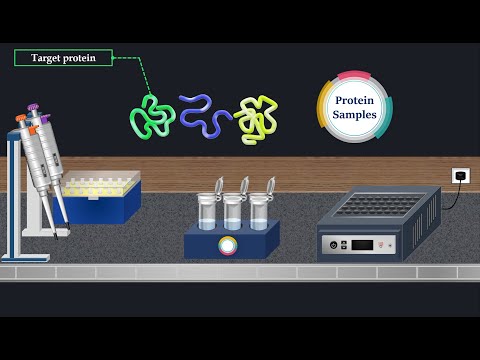
Introduction to Western Blotting
- Western blotting is a crucial method in cell and molecular biology for protein separation and detection, allowing the identification of specific proteins from complex mixtures like cell lysates.
- The technique involves three main steps: separation by size, transfer of proteins to a solid support, and detection using antibodies.
SDS-PAGE Procedure
- Sample preparation begins with adding a loading buffer containing SDS, beta-mercaptoethanol, bromophenol blue, and glycerol to ensure uniform negative charge across all proteins.
- Samples are heated to 95°C; this denatures proteins by disrupting non-covalent interactions such as hydrogen bonds and ionic bonds.
- SDS (anionic surfactant) binds uniformly to proteins, making their intrinsic charges negligible compared to the negative charges from SDS.
Gel Preparation and Electrophoresis
- A polyacrylamide gel is created between two glass plates; electrodes are positioned with the positive electrode at the bottom.
- During electrophoresis, a molecular-weight size marker is loaded onto the gel for estimating protein sizes based on migration patterns.
- An electric field causes negatively charged proteins to migrate towards the positive electrode; smaller proteins move faster through the gel matrix.
Stopping Electrophoresis
- The migration can be visually monitored using bromophenol blue dye which migrates faster than most proteins; this allows stopping before complete migration occurs.
Protein Transfer Process
- After separation, proteins are transferred from the gel onto a membrane using wet transfer methods involving various layers including blotting paper and fiber pads.
- Nitrocellulose or PVDF membranes are used for transferring separated proteins while maintaining their organization from the gel.
Electroblotting Methodology
- Electroblotting is commonly employed for transferring proteins. It uses an electric current to pull negatively charged proteins onto positively charged membranes.
Western Blotting Process Overview
Steps in Protein Detection
- The process begins with the removal of each layer of the fiber pad until reaching the membrane, followed by electrotransfer of proteins onto the membrane.
- A blocking solution containing bovine serum albumin (BSA) is applied to prevent non-specific interactions between the membrane and antibodies used for detecting target proteins.
- After incubation, the blocking solution is removed, and a primary antibody solution is added. This antibody specifically binds to the target protein on the membrane.
- Following incubation with the primary antibody, unbound antibodies are washed away using a wash buffer to minimize background noise in detection.
Secondary Antibody Application
- A secondary antibody solution is introduced after washing; this antibody recognizes and binds to a specific portion of the primary antibody.
- Thorough washing of the membrane at this stage is crucial to remove any unbound secondary antibodies before proceeding to detection.
Chemiluminescent Detection Method
- The membrane is incubated in a substrate solution for chemiluminescent detection, commonly linked with horseradish peroxidase (HRP).
- In presence of hydrogen peroxide, HRP catalyzes oxidation of luminol, producing light at 425 nm proportional to HRP-conjugated secondary antibodies and indirectly measuring target protein presence.
Final Analysis