Chapter 13 Central Nervous System
Chapter 13: The Central Nervous System
Overview of the Lecture
- Introduction to Chapter 13, focusing on the central nervous system (CNS), specifically the brain and spinal cord.
- Discussion on embryological development of the brain to understand its regions better.
- Emphasis on the cerebral cortex as a key area for processing information and intelligence.
- Overview of protective structures like meninges and their continuity with the spinal cord.
- Clarification that CNS consists solely of the brain and spinal cord, highlighting their control functions.
Functions of the Brain
- The brain acts as a control center for vital functions such as heart rate, respiratory rate, and blood pressure.
- Mention of autonomic nervous system and endocrine system's relationship with brain control via hypothalamus.
- Complex neural functions including intelligence, consciousness, memory, emotion, behavior, and socialization are centered in the brain.
- Sensory information is processed in specific areas of the brain before motor responses are sent out through peripheral nerves.
Embryonic Development of the Brain
- Introduction to directional terms: "rostral" (towards nose) and "caudal" (towards tail).
- Explanation that the brain develops from the rostral part of the neural tube during early embryonic stages.
Primary Brain Vesicles
- Identification of three primary vesicles: prosencephalon (forebrain), mesencephalon (midbrain), rhombencephalon (hindbrain).
Secondary Brain Vesicles
- After five weeks, primary vesicles divide into secondary ones; prosencephalon divides into telencephalon and diencephalon while mesencephalon remains undivided.
- Rhombencephalon divides into metencephalon and myelencephalon; emphasis on retaining "diencephalon" name for important adult structures.
Adult Structures Derived from Secondary Vesicles
- Telencephalon becomes cerebrum or cerebral hemispheres—largest portion of brain mass.
Brain Development and Structure
Overview of Brain Regions
- The mesencephalon, metencephalon, and myelins f1 develop into parts of the brainstem: midbrain, pons, and medulla oblongata. The metencephalon also forms the cerebellum.
- Rapid growth of cerebral hemispheres envelops the diencephalon and midbrain regions, resembling a mushroom cap over the brainstem. This growth leads to wrinkling due to limited skull space.
- Enlargements in the neural tube create ventricles filled with cerebrospinal fluid (CSF), which develop as the brain grows rapidly.
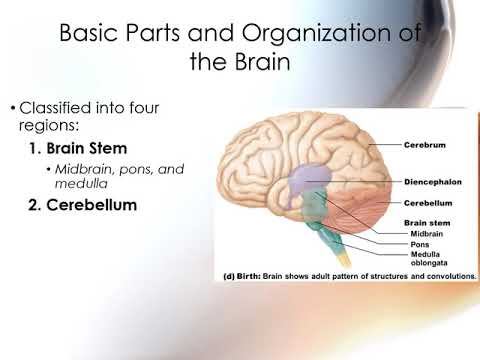
- At birth, four main regions are established: brainstem (midbrain, pons, medulla), cerebellum ("little brain"), diencephalon (thalamus, hypothalamus, epithalamus), and cerebrum (two hemispheres).
Organization of Brain Matter
- Gray matter contains neuron cell bodies while white matter consists of axons forming tracts. In the brain's structure, gray matter is centrally located with white matter surrounding it.
- The outermost layer of the cerebrum and cerebellum is called the cortex or cerebral cortex; this layer is formed from migrating cell bodies.
- Unlike other areas like the spinal cord where gray matter is central and white matter external, only the cerebrum and cerebellum have an external gray matter layer.
Ventricular System
- Ventricles are expansions of the neural tube's central cavity filled with CSF. Ependymal cells help circulate this fluid around the brain and spinal cord.
- Choroid plexus within each ventricle produces CSF through filtration from blood capillaries.
- All ventricles are interconnected; they connect to each other as well as to the central canal in the spinal cord for continuous flow of CSF.
Detailed Structure of Ventricles
- There are four ventricles: lateral ventricles shaped like sheep horns that extend into cerebral hemispheres; third ventricle located between them; fourth ventricle connects to central canal via cerebral aqueduct.
- Visual aids can enhance understanding of ventricular locations within a three-dimensional model of the brain.
This structured overview captures key insights about brain development stages, organization by type of tissue (gray vs. white matter), and details on how cerebrospinal fluid circulates through various structures in developing brains.
Brain Stem Anatomy and Functions
Overview of the Brain Stem
- The brain stem consists of three main parts: the midbrain (most rostral), pons (middle), and medulla oblongata (caudal). It serves as a passageway for fiber tracts, primarily axons connecting the cerebrum to the spinal cord.
- Fiber tracts are crucial for neuronal communication between the cerebrum and spinal cord, facilitating signals that travel through the brain stem before reaching spinal nerves.
Cranial Nerves and Autonomic Functions
- Ten out of twelve cranial nerves originate in the brain stem, playing significant roles in various bodily functions. Further details on these nerves will be discussed in subsequent lectures.
- The brain stem is responsible for involuntary or autonomic behaviors essential for survival, such as heart rate and respiratory control.
Medulla Oblongata
- The medulla oblongata connects directly with the spinal cord and features external landmarks like pyramids where motor tracks cross over (decussation).
- Decussation explains why each side of the brain controls opposite sides of the body; this crossover occurs at specific points within the medulla.
- Cerebellar peduncles connect different parts of the brain; there are three types associated with linking cerebellum to both cerebrum and brain stem.
Functions of Medulla Oblongata
- Four pairs of cranial nerves emerge from the medulla, including important ones like cranial nerve 10 (vagus nerve).
- The core contains reticular formation involved in autonomic functions regulating visceral organs such as cardiovascular and respiratory centers.
Pons
- The pons acts as a bridge between midbrain and medulla oblongata, housing three cranial nerves related to facial sensations/movements and eye movements.
- It plays a vital role in motor skills by connecting cerebral cortex signals to cerebellum via middle cerebellar peduncle.
Midbrain
- Positioned between diencephalon and pons, it contains cerebral aqueduct connecting third and fourth ventricles.
- Distinct from cerebellar peduncles, cerebral peduncles link midbrain to cerebrum on its ventral surface.
Cerebral Structures and Functions
Overview of the Midbrain
- The cerebrum connects to the midbrain and brain stem, with notable nuclei present in the midbrain, including dorsal and ventral nuclei.
- The corpora quadrigemina serve as a landmark in the midbrain, consisting of two superior colliculi (involved in visual reflexes) and two inferior colliculi (involved in auditory reflexes).
- The red nuclei are significant ventral nuclei that play a role in the reticular formation, which is crucial for autonomic functions.
Introduction to the Cerebellum
- The cerebellum, referred to as the "little brain," is located dorsal to the brain stem and has a similar shape to the cerebrum.
- It consists of two hemispheres with folds known as folia; a structure called vermis lies between these hemispheres.
Structure of the Cerebellum
- Composed of three regions: an outer cortex (gray matter), internal white matter known as arbor vitae, and some deep nuclei.
- The longitudinal section reveals white matter resembling a tree structure surrounded by folia.
Functions of the Cerebellum
- Primarily involved in smoothing and coordinating body movements while maintaining equilibrium; it receives movement information from the motor cortex.
- After deciding on a movement, the motor cortex sends this information to the cerebellum for fine-tuning before executing it.
Disorders Related to Cerebellar Function
- An example includes cerebellar hypoplasia in cats, where underdevelopment leads to intention tremors during movement execution.
Higher Cognitive Functions of the Cerebellum
Learning and Cognition
- Besides motor function coordination, it plays roles in learning new motor skills and higher cognitive tasks such as language processing and problem-solving.
Communication Pathways
- The cerebellum communicates with other brain structures via cerebellar peduncles connecting it to various parts of the brainstem.
Exploring Diencephalon Structures
Components of Diencephalon
- The diencephalon serves as a central core within the forebrain comprising three main structures: thalamus (largest), hypothalamus (below thalamus), and epithalamus (smallest).
Diencephalon and Its Functions
Overview of the Diencephalon
- The diencephalon serves as a critical border for the third ventricle, positioned between the left and right thalamus.
- The thalamus can be viewed as two hemispheres connected by the interthalamic adhesion, which is located centrally in front of the thalamus.
Structure and Function of Thalamus
- The third ventricle is a circular structure that encircles the interthalamic adhesion, acting as a boundary for sensory information processing.
- Comprising about 80% of the diencephalon, the thalamus contains numerous nuclei that function primarily as relay stations for incoming sensory data.
- It directs sensory information to specific regions of the cerebral cortex, akin to an old-school switchboard managing calls.
Sensory Information Processing
- The thalamus determines whether sensory signals are significant or trivial; it amplifies important signals while downplaying less critical ones.
- This amplification or attenuation process is managed by various nuclei within the thalamus.
Hypothalamus: Control Center
Role in Autonomic Nervous System
- Positioned rostrally to the thalamus, the hypothalamus plays a vital role in regulating both autonomic functions and endocrine activities.
- It lies between key structures such as the optic chiasm and mammillary bodies, with many nuclei contributing to its functions.
Functions of Hypothalamus
- Acts as a primary visceral control center influencing body temperature, hunger, thirst, emotional responses, sleep-wake cycles, and more.
- It also has connections to memory formation but is chiefly recognized for its regulatory roles over autonomic and endocrine systems.
Epithalamus: Pineal Gland's Role
Structure and Function
- The epithalamus forms part of the roof of the third ventricle and includes several small nuclei like the pineal gland.
- The pineal gland produces melatonin which regulates circadian rhythms—essential for sleep-wake cycles.
Cerebral Hemispheres: Overview
Characteristics of Cerebrum
- Representing 83% of brain mass, cerebral hemispheres grow rapidly leading to folding that increases surface area through grooves known as fissures.
- Major deep grooves separate different brain regions; one significant fissure is the transverse fissure separating cerebrum from cerebellum.
Understanding the Structure of the Cerebrum
Overview of Brain Anatomy
- The brain is divided into two main parts: the cerebrum and cerebellum, separated by the longitudinal fissure, which runs down the middle of the head.
- The surface of the cerebrum features grooves called sulci (singular: sulcus) and ridges known as gyri (singular: gyrus), which are consistent across individuals due to their representation in the cerebral cortex.
Lobes of the Cerebral Cortex
- The lobes of the cerebral cortex are named after overlying skull bones: frontal lobe (under frontal bone), parietal lobe (under parietal bones), occipital lobe (under occipital bone), and temporal lobes (on either side).
- A deeper region called the insula lies beneath the temporal lobe, visible when separating it from both frontal and temporal lobes.
Important Sulci and Gyri
- The central sulcus is a key landmark that separates frontal and parietal lobes; it has significant gyri on either side—pre-central gyrus (motor functions) and post-central gyrus (sensory functions).
- Other notable sulci include:
- Parieto-occipital sulcus, separating occipital from parietal lobe.
- Lateral sulcus, separating temporal lobe from both parietal and frontal lobes; opening this reveals the insula.
Functions of the Cerebral Cortex
- The cerebral cortex is crucial for conscious thought, self-awareness, memory storage, voluntary movement control, communication through language and facial expressions—all housed within its gray matter.
- Comprising about 40% of total brain mass, its folds allow for increased size without expanding overall volume.
Studying Functional Areas
- Modern imaging techniques like PET scans and MRIs have replaced older methods for studying brain function. These tools help identify distinct regions responsible for motor and sensory activities.
- The cerebral cortex can be categorized into functional areas:
- Primary sensory cortices correspond to specific senses (e.g., visual or auditory).
- Sensory association areas process information from primary sensory cortices.
- Multimodal association areas integrate multiple senses simultaneously.
Motor Function Areas
- There exists a motor association area known as pre-motor cortex that plans movements alongside a singular primary motor cortex responsible for executing these actions.
Understanding Sensory and Motor Areas of the Brain
Overview of Sensory Areas
- The brain's sensory areas are organized by type, including sensory (taste, vision) and motor functions, with multi-modal association areas integrating various senses.
- Primary sensory areas for taste, vision, hearing, and touch are located in specific regions; large pink areas represent multi-modal association zones.
- Sensory information is relayed through the thalamus to the corresponding primary sensory cortex before reaching association areas for further processing.
Processing Information
- After initial processing in primary sensory cortices, information moves to multimodal association areas for parallel processing before being sent to motor cortices.
- Major sensory cortices include those for somatosensory (touch), visual (largest in humans), auditory (sound), vestibular (balance), olfactory (smell), gustatory (taste), and visceral senses.
Motor Areas of the Brain
- Motor areas are primarily located in the posterior frontal lobe; they control voluntary movements based on processed sensory information.
- The primary motor cortex governs somatic motor functions while the pre-motor cortex plans complex movements and includes specialized regions like the frontal eye field and Broca's area.
Speech Production and Emotional Tone
- The left hemisphere is responsible for speech production while the right hemisphere manages emotional nuances in speech delivery.
- This division highlights distinct functional roles between hemispheres regarding language processing.
Body Mapping: Somatosensory vs. Motor Cortex
- A body map illustrates both somatosensory and primary motor cortices' locations relative to the central sulcus; it emphasizes their roles in sensation and movement control.
- The post-central gyrus corresponds to the primary somatosensory cortex while the pre-central gyrus relates to primary motor functions, showcasing their anatomical significance.
Understanding the Cerebral Cortex and Its Functions
Overview of Body Mapping in the Cortex
- The body map illustrates different regions of the body, with larger areas indicating greater fine motor control or sensation.
- This mapping highlights various areas along the gyri and sulci of the cortex, emphasizing their significance in sensory processing.
Multimodal Association Areas
- The cerebral cortex contains multimodal association areas that process information from multiple sensory modalities, facilitating complex associations.
- There are three key multimodal association areas: posterior association area, anterior association area (prefrontal cortex), and limbic association area located medially.
Posterior Association Area
- Positioned between visual, auditory, and somatosensory areas, it aids in spatial awareness of body locations.
- It integrates visual and auditory information to identify objects' locations in space as well as one's own body position.
Anterior Association Area (Prefrontal Cortex)
- This large area is crucial for integrating past experiences with current sensory input to plan motor movements.
- It governs complex functions such as reasoning, judgment, social skills, impulse control, and mental flexibility.
Developmental Insights on Prefrontal Cortex
- The prefrontal cortex matures last during adolescence; its underdevelopment contributes to impulsive decision-making typical in teenagers.
- It retains information for only 30 seconds but interacts with memory systems through the limbic system.
Limbic System Functionality
- Located medially within the cerebral hemispheres, it plays a vital role in memory formation and emotional processing.
- The limbic system integrates sensory inputs with motor behaviors to help formulate memories and manage emotions.
Contralateral Control of Hemispheres
- Each hemisphere controls opposite sides of the body; left hemisphere excels at language and logic while right hemisphere specializes in visual-spatial skills and emotional understanding.
- Lateralization indicates that cognitive functions are distributed unevenly across hemispheres; individuals may exhibit strengths on one side over another.
Communication via Cerebral White Matter
- Cerebral white matter facilitates communication among different cortical areas as well as between the brain stem and spinal cord.
Understanding White Matter Tracts in the Brain
Overview of Myelinated Axons
- Myelinated axons are grouped into tracts, which bundle together to connect specific areas of the brain. The coronal section of the brain shows both gray matter (cortex) and white matter (tracts).
Types of White Fiber Tracts
- There are three types of white fiber tracts: commissural fibers, association fibers, and projection fibers.
- Commissural Fibers: Facilitate communication between the two cerebral hemispheres; prominently represented by the corpus callosum.
- Association Fibers: Connect different parts within the same hemisphere, linking various cortical areas.
- Projection Fibers: Descend from the cerebral cortex to lower body regions, including pathways through the brainstem and spinal cord.
Contralateral Control Mechanism
- The medulla oblongata is crucial for contralateral control where axons cross over; right-side axons influence left-side body functions and vice versa.
Protective Structures Surrounding the Brain
Meninges and Their Functions
- The skull protects the brain physically, while meninges serve as connective tissue layers that encase it. They also protect blood vessels supplying the brain.
Layers of Meninges
- There are three layers:
- Dura Mater: Outermost layer; strongest with two sub-layers containing dural sinuses for venous drainage.
- Arachnoid Mater: Middle layer housing cerebrospinal fluid (CSF); named for its spider-web-like appearance due to arachnoid trabeculae.
- Pia Mater: Innermost delicate layer closely adherent to brain surface.
Cerebrospinal Fluid (CSF)
- CSF provides cushioning for protection, nourishes neural tissues with nutrients from blood, and removes waste products. It circulates through ventricles and around the brain.
Formation and Circulation of CSF
- Produced in choroid plexus located in each ventricle; involves capillary beds and ependymal cells facilitating nutrient transfer from blood to CSF.
Understanding Cerebrospinal Fluid and the Blood-Brain Barrier
Cerebrospinal Fluid Production and Flow
- Cerebrospinal fluid (CSF) is produced in the choroid plexus located within the ventricles of the brain. It flows through all ventricles and the central canal of the spinal cord, as well as through the subarachnoid space between meninges.
- CSF is absorbed into the dural sinus via arachnoid villi, which are small projections that facilitate this process.
The Blood-Brain Barrier
- The blood-brain barrier (BBB) is primarily associated with the choroid plexus and consists of impermeable capillaries that prevent blood-borne toxins from entering cerebral spinal fluid or brain tissue.
- While it restricts many substances, it allows essential molecules like oxygen and glucose to pass through. Additionally, certain substances such as alcohol, nicotine, and anesthetics can cross this barrier.
Overview of Spinal Cord Anatomy
Structure and Function
- The spinal cord runs through the vertebral canal from the foramen magnum at the base of the skull to approximately L1-L2 vertebrae where it terminates.
- It serves as a two-way conduction pathway for sensory information traveling to the brain and motor commands traveling to various body parts. Reflex centers are also located here.
Anatomical Features
- The spinal cord has two deep grooves: an anterior fissure (deeper groove) and a posterior median sulcus, giving it a butterfly appearance.
- The termination point of the spinal cord is known as conus medullaris; below this point lies cauda equina, which consists of nerve roots extending downwards.
Spinal Cord Cross Section Analysis
Gray Matter vs. White Matter
- In contrast to brain anatomy, white matter in the spinal cord is located on its exterior while gray matter forms an internal "H" or butterfly shape.
- White matter contains myelinated axons facilitating communication between different regions of the spinal cord and brain; some non-myelinated axons are also present.
Functional Regions
- Gray matter comprises cell bodies with dorsal horns receiving sensory input from peripheral nerves while ventral/lateral horns send motor signals outwards.
- A connection exists between left and right sides called gray commissure; additionally, a central canal filled with cerebrospinal fluid runs longitudinally through this region.
Understanding the Spinal Cord and Central Nervous System
Overview of Sensory and Motor Pathways
- The spinal cord integrates sensory information from the body, with sensory neurons entering through the dorsal (posterior) roots.
- Dorsal root ganglions house the cell bodies of these sensory neurons; motor output exits via the ventral root containing motor neurons.
- Dorsal roots are purely sensory, while ventral roots are purely motor; together they form a spinal nerve that carries both types of signals.
Meninges and Cerebrospinal Fluid
- The spinal cord is protected by three layers of meninges: dura mater, arachnoid mater, and pia mater, along with an epidural space filled with fat.
- Epidurals target this epidural space to deliver anesthetics or anti-inflammatories affecting spinal nerve roots.
- Cerebrospinal fluid flows through the central canal and subarachnoid space, connecting brain and spinal cord.
Ascending and Descending Pathways
- In ascending pathways (sensory), signals must pass through the thalamus before reaching their destination in the cortex.
- Descending pathways originate from the primary motor cortex; they cross over at the medulla oblongata before traveling down to muscles via ventral roots.
Disorders of the Central Nervous System
Spinal Cord Damage
- Effects of spinal cord damage depend on location; paralysis relates to motor function while paresthesia pertains to sensory function.
- Paraplegia affects lower limbs due to thoracic or lumbar damage; quadriplegia results from cervical spine injury.
Brain Dysfunction
- Concussions involve brain jarring leading to temporary effects; repeated concussions can cause long-term issues seen in sports like football.
- A cerebrovascular accident (stroke) disrupts blood flow to the brain, risking neuron death after five minutes without oxygen.
Progressive Degenerative Diseases
- Alzheimer's disease leads to memory loss and confusion due to progressive degeneration primarily affecting cortical areas.
- Hydrocephalus involves excessive cerebrospinal fluid causing skull enlargement; neural tube defects can lead to severe conditions like spina bifida.
Understanding Spinal Malformations and Cerebral Palsy
Spinal Malformations
- The vertebrae can be malformed, leading to issues with the vertebral arch and canal, which may cause spinal fluid leakage.
- This condition can result in myelomeningoceles, a severe form of spina bifida characterized by an open spinal cord.
- Myelomeningoceles can have detrimental effects on health but are often correctable through surgery if not too severe.
Cerebral Palsy
- Cerebral palsy is described as a voluntary muscle control issue due to damage to the motor cortex.
- The damage may arise from either an injury or malformation of the brain, affecting voluntary control over skeletal muscles.
- This condition highlights the importance of understanding neurological impacts on muscle function.