ESTRUTURA E FUNÇÃO DA MEDULA ESPINAL - PARTE 2
Medullary Structure and Descending Pathways
Overview of Spinal Cord Structure
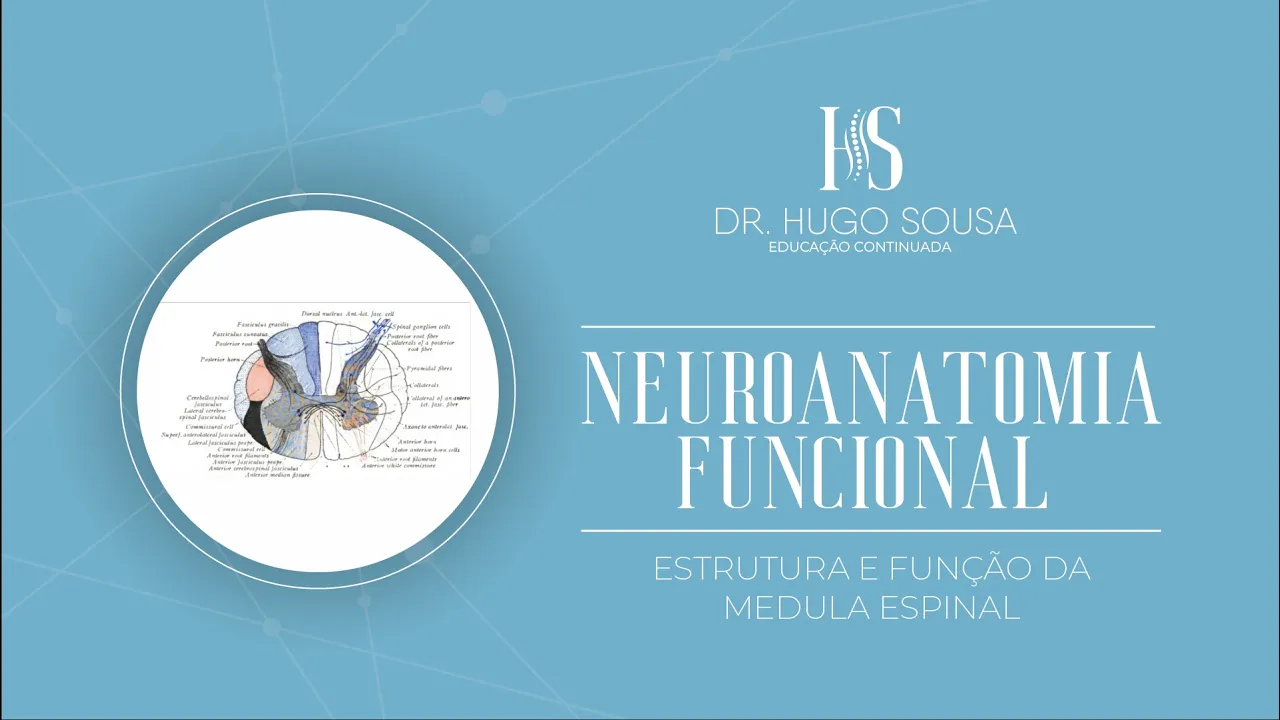
- The spinal cord is divided into white matter segments known as funiculi, which are synonymous with cords. There are four main funiculi: anterior, lateral, and posterior.
Ascending and Descending Tracts
- Funiculi contain tracts and fascicles; red represents descending motor tracts while blue indicates ascending sensory tracts.
Introduction to Descending Pathways
- The discussion shifts to descending pathways, highlighting their complexity and connections with the anterior column of the spinal cord.
Key Descending Tracts
- Important descending tracts include:
- Lateral corticospinal tract (vital for motor function)
- Anterior corticospinal tract
- Rubrospinal tract
- Vestibulospinal tract
- Reticulospinal tract
- Tectospinal tract
Lateral Corticospinal Tract Details
- The lateral corticospinal tract originates from the cerebral cortex, specifically from pyramidal cells, projecting downwards through the internal capsule to connect with the anterior portion of the spinal cord.
Corticospinal Tract Pathway
Pathway Description
- This pathway descends from higher brain centers to lower levels, passing through critical structures like the internal capsule and midbrain peduncles before reaching the medulla.
Decussation of Fibers
- Most fibers in this tract cross at the medullary pyramids; those on the right side will project to the left side of the spinal cord's lateral funiculus.
Clinical Implications of Lesions
Effects of Lesions Above Decussation
- Lesions occurring above decussation (e.g., in cortex or internal capsule) result in contralateral motor symptoms due to uncrossed fibers still projecting downward.
Effects of Lesions Below Decussation
- Conversely, lesions below decussation lead to ipsilateral symptoms since affected fibers have already crossed over within the spinal cord.
Functionality of Corticospinal Tract
Motor Control Role
- The primary function is voluntary control over distal musculature, particularly fine motor skills essential for precision tasks.
Rubrospinal Tract Overview
Origin and Functionality
Understanding Brainstem Injuries and Motor Responses
Manifestation of Brainstem Injuries
- Different levels of the brainstem exhibit varied manifestations depending on the type of injury; localized injuries show distinct motor responses compared to diffuse injuries.
- If the brainstem is preserved, motor activity patterns differ when only the cortex is disconnected from other body parts and nervous system areas.
Medial System Overview
- Introduction to the medial system, focusing on specific fiber tracts involved in motor control.
- The anterior corticospinal tract originates in the cortex and targets the spinal cord's anterior column without crossing at the medulla's pyramidal decussation.
Crossing Patterns of Motor Fibers
- Most fibers (75%-80%) cross at the pyramidal decussation, while 20%-25% cross at segmental levels within the spinal cord.
- This crossing pattern is crucial for understanding voluntary motricity related to axial muscles and proximal upper limb muscles.
Tecto-Spinal Tract Functionality
- The tecto-spinal tract originates from regions linked to sensory-motor orientation, particularly from superior colliculi in the midbrain.
- It facilitates reflexive head movements in response to auditory or visual stimuli, allowing quick reactions to environmental changes.
Reticulo-Spinal Tract Insights
- The reticulo-spinal tract arises from a network of neurons known as the reticular formation located throughout the brainstem.
- This formation plays a vital role in maintaining life-sustaining reflexes due to its extensive connections across various brainstem nuclei.
Postural Adjustments and Motor Control
- The pontine reticulo-spinal tract aids postural adjustments by activating extensor muscles in lower limbs while also influencing voluntary motricity similar to anterior corticospinal functions.
Understanding the Role of Muscles in Breathing and Posture
The Automatic Function of the Diaphragm
- The diaphragm operates automatically during breathing, highlighting its essential role without conscious effort from individuals.
- While we can alter our breathing patterns, the primary function remains involuntary, emphasizing the body's natural respiratory mechanisms.
Muscle Groups and Voluntary Motor Control
- Striated muscles in the axial region are directly linked to voluntary motor functions, indicating their importance in controlled movements.
- The medial grouping known as lateral and medial vestibulospinal tracts is crucial for postural adjustments and maintaining balance.
Vestibular Nuclei and Balance Maintenance
- Vestibular nuclei located in the pons connect with the cerebellum, playing a significant role in processing information related to posture and balance.
- These nuclei receive sensory input from Golgi tendon organs and muscle spindle fibers, which are vital for proprioception.
Muscle Tone Adjustments
- Information regarding muscle tone adjustments is critical for rapid responses to changes in posture or movement speed.
- The cerebellum facilitates quick reflexive responses to maintain balance when unexpected disturbances occur.
Cerebellar Functionality in Response to Imbalance
- The cerebellum processes information from the spinal cord reflexively, allowing for immediate adjustments without waiting for cortical decision-making.