🥇 Anatomía de la PLEURA. Fácil, Rápida y Sencilla.
Anatomy of the Pleura
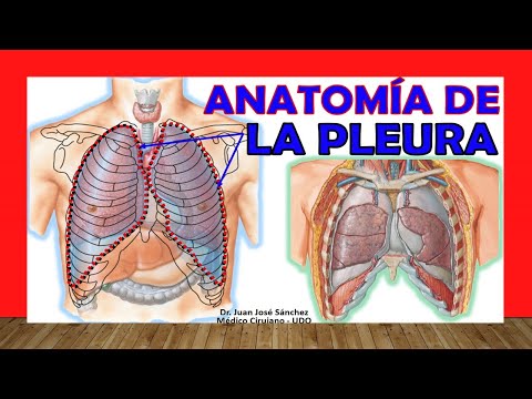
In this section, Juan José Sánchez introduces the topic of the anatomy of the pleura, focusing on its structure and functions in relation to the lungs.
Anatomy of the Pleura
- The pleura is a serous membrane that covers and protects the lungs. It consists of two main parts: visceral pleura (not easily found in atlases as it is part of the lung itself) and parietal pleura (visible in atlases and related to the walls of the thoracic cavity).
- Understanding the cavities within which these structures lie is crucial for grasping pleural anatomy. Mastery of thoracic cavity components like diaphragm, sternum, and ribs aids in comprehending pleural structures.
- The space between visceral and parietal pleura forms a virtual cavity known as the pleural cavity. This space contains pleural fluid that facilitates smooth movement during breathing by reducing friction between layers.
Layers and Divisions
- The layers from superficial to deep include: thoracic wall, intercostal muscles, endothoracic fascia, parietal pleura, pleural cavity, visceral pleura, and subpleural tissue. Understanding these layers aids in visualizing their relationships within the thorax.
Anatomy of the Thoracic Wall
In this section, the speaker discusses the anatomy of the thoracic wall, focusing on the pleura and its different components.
Understanding the Anatomy of the Thoracic Wall
- The thoracic wall consists of structures like the vertebral column (posterior), sternum (anterior), and ribs that connect these two structures.
- Differentiating between pleura parietal mediastinica (facing towards mediastinum) and pleura parietal costal (in contact with sternum, ribs, and part of vertebral column).
- Identifying pleura parietal diafragmatica (facing downwards towards base) and understanding its location in relation to other components like pleura parietal costal.
Understanding Pleural Borders
This section delves into the borders of the pleura and their significance in connecting different parts of the thoracic cavity.
Exploring Pleural Borders
- Examining how different borders unite portions of the pleura, such as inferior border linking cúpula pleural with mediastinal and diaphragmatic pleura.
- Discussing posterior border responsible for connecting mediastinal with costal parietal pleura near or at vertebral column level.
- Highlighting anterior border as a crucial connection point between mediastinal and costal parietal pleura anteriorly, close to posterior aspect of sternum.
Recesses in Pleural Cavity
This segment focuses on recesses within the pleural cavity, exploring spaces where visceral pleura transitions to parietal pleura.
Unveiling Pleural Recesses
- Introducing concepts of recesos or senos as cavities within the pleural space not typically occupied by lung tissue.
- Identifying specific locations like costo-mediastinal recess between mediastinal and costal pleura, aiding medical professionals in diagnostic processes.
- Exploring costo-diaphragmatic recess situated between diaphragmatic and costal surfaces, providing insights into anatomical relationships within thoracic cavity.
Anatomy of the Pleura: Irrigation, Venous Drainage, and Innervation
In this section, the speaker discusses the anatomy of the pleura focusing on irrigation, venous drainage, and innervation. The distinction between visceral and parietal pleura is highlighted along with their respective vascular supplies and nerve innervations.
Vascular Supply to Parietal Pleura
- The parietal pleura receives dense irrigation from:
- Posterior intercostal arteries (branches of thoracic aorta)
- Superior intercostal arteries (branches of subclavian artery)
- Internal mammary arteries (supplying anterior intercostal arteries)
- Phrenic or superior diaphragmatic arteries (branches of aorta)
Vascular Supply to Visceral Pleura
- The visceral pleura lacks direct vascular supply from thoracic cage structures but is irrigated by bronchial arteries (aortic branches).
Venous Drainage
- Venous drainage differs for parietal and visceral pleura:
- Parietal pleura: Drained by posterior and superior intercostal veins, internal mammary veins, and azygos system.
- Visceral pleura: Drained by pulmonary veins.
Innervation of Parietal Pleura
- Parietal pleura is highly sensitive to pain due to dense innervation:
- Costal part: Innervated by intercostal nerves
- Diaphragmatic part: Innervated by phrenic nerve medially and subcostal nerve laterally
- Mediastinal part: Innervated by phrenic nerve
Contrasting Sensitivity
- While parietal pleura is sensitive to pain due to rich innervation causing pleuritic pain, visceral pleura lacks such sensitivity as it is part of the lung tissue.