Inflamação, Resposta inflamatória, Quimiocinas; Migração leucocitária; Fagocitose.
Introduction to Inflammation
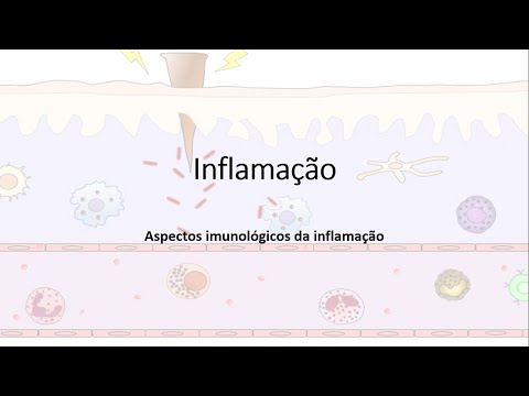
The instructor introduces the topic of inflammation and explains that in this immunology class, they will focus on the immunological aspects of inflammation.
Understanding Inflammation
- Inflammation is a response of the immune system to eliminate pathogens or damaged tissues.
- It can occur due to infection, tissue damage, or autoimmune diseases.
- Autoimmune diseases result from the immune system attacking its own cells and tissues.
Types of Inflammation
The instructor discusses different types of inflammation and their significance.
Autoimmune Inflammation
- Autoimmune diseases can lead to inflammation when the immune system mistakenly targets self-cells and tissues.
- Examples include systemic lupus erythematosus (SLE), where antibodies attack nuclear material and proteins in cells.
Other Causes of Inflammation
- Inflammation can also be triggered by infections, physical or chemical agents, and environmental substances.
- Microorganisms, foreign bodies like splinters, burns, radiation exposure, and chemical substances can all induce inflammation.
Effects of Inflammation
The instructor explains the effects of inflammation on tissues.
Changes in Blood Vessels
- During inflammation, blood vessels may dilate (increase in diameter) and become more permeable.
- This allows increased blood flow to the affected area and facilitates the entry of immune cells into tissues.
Visualizing Normal vs. Inflamed Tissues
The instructor compares normal and inflamed tissues visually to highlight differences during inflammation.
Comparison of Normal Tissue vs. Inflamed Tissue
- When comparing normal tissue to inflamed tissue, the inflamed tissue shows a larger vessel caliber and increased thickness.
- These visual differences indicate the presence of inflammation in the tissue.
The transcript does not provide further sections or timestamps for additional topics.
Increase in Blood Flow and Permeability
In this section, the speaker discusses the increase in blood flow and permeability due to the dilation of blood vessels. They explain how this leads to an increase in blood pressure and the accumulation of plasma in the inflamed tissue.
Increase in Blood Flow
- The increase in caliber of blood vessels leads to a higher volume of blood being directed towards the inflamed area. This results in an increased blood flow.
Permeability of Blood Vessels
- The speaker uses an analogy with rainfall to explain permeability. In areas with impermeable surfaces like asphalt, rainwater cannot penetrate, while in areas with permeable soil and trees, water can enter the ground.
- Increased permeability allows components of the blood, such as plasma, to easily move from inside the vessels to outside into the surrounding tissue.
- This increased permeability is one of the effects of inflammation.
Effects on Tissue
- The increased blood flow and permeability result in several effects on tissue:
- Accumulation of plasma (the liquid part of blood) in the inflamed area.
- Changes in cell composition, with more cells present compared to normal tissue.
- These cells are responsible for migration from inside blood vessels to outside during inflammation.
- This migration is known as leukocyte migration or leukocytic diapedesis.
Inflammatory Signs: Calor, Rubor, Tumor, Dolor
The speaker explains four cardinal signs of inflammation: calor (heat), rubor (redness), tumor (swelling), and dolor (pain). They discuss how these signs are related to vasodilation, increased metabolism, accumulation of plasma, and the release of inflammatory mediators.
Calor (Heat)
- Vasodilation during inflammation leads to an increase in local temperature.
- This is due to the higher metabolic activity in the area.
Rubor (Redness)
- Vasodilation causes increased blood flow, resulting in redness in the inflamed area.
- The increased blood flow brings more oxygen and nutrients to the tissue.
Tumor (Swelling)
- Swelling occurs due to the accumulation of plasma and other fluids outside the blood vessels.
- Increased permeability allows these components to move from inside vessels to outside into the surrounding tissue.
Dolor (Pain)
- Inflammatory mediators released during inflammation can stimulate nerve endings, leading to pain.
- Nerve endings are present near blood vessels and are sensitive to these mediators.
- The release of chemical mediators triggers pain signals through nerve terminals.
Loss of Function as a Sign of Inflammation
The speaker explains that loss of function is another sign of inflammation. They mention that this loss is temporary and that once inflammation subsides, normal tissue function is restored.
Loss of Function
- During inflammation, there may be a temporary loss or impairment of normal tissue function.
- Once the inflammatory process resolves, tissue function returns to normal.
Inflammatory Mediators: Lipids and Chemical Mediators
The speaker discusses inflammatory mediators released during inflammation. They explain that lipids derived from arachidonic acid play a role in initiating inflammation. They also mention chemical mediators, such as histamines and cytokines, which are released by mast cells.
Lipid Mediators
- Inflammatory mediators derived from arachidonic acid play a role in initiating inflammation.
- These lipid mediators are released from the plasma membrane of mast cells.
Chemical Mediators
- Mast cells are the main producers of inflammatory mediators, including histamines and cytokines.
- These chemical mediators are substances released by cells to promote or regulate inflammation.
Overview of Phospholipase and Arachidonic Acid Pathways
This section provides an overview of the pathways involving phospholipase and arachidonic acid.
Phospholipase and Arachidonic Acid Pathways
- Phospholipase releases arachidonic acid from phospholipids.
- Arachidonic acid is processed by two main pathways: lipoxigenases and ciclooxigenases.
- Lipoxigenases pathway leads to the production of leukotrienes, which can cause cell migration to specific regions.
- Ciclooxigenases pathway leads to the production of prostaglandins, thromboxanes, and other mediators involved in vasodilation, edema, vasoconstriction, and bronchial constriction.
Anti-inflammatory Drugs
This section discusses anti-inflammatory drugs and their effects on the inflammatory response.
Corticosteroids
- Corticosteroids are potent anti-inflammatory drugs that directly inhibit phospholipase enzyme activity.
- They prevent the processing of arachidonic acid into inflammatory mediators.
- Corticosteroids should be used under medical guidance due to potential side effects.
Other Anti-inflammatory Drugs
- H2F lysalpha bife, prostaglandins, tromboxanes, and other mediators are also involved in inflammation.
- Inhibitors of COX1 and COX2 are less potent anti-inflammatory drugs compared to corticosteroids.
Chemokines in Immune Response
This section explains the role of chemokines in immune responses.
Chemokines as Cytokines
- Chemokines are a type of cytokine involved in immune responses.
- They control cell adhesion and chemotaxis, which is the movement of cells to specific locations based on chemokine concentration.
Cell Movement and Chemotaxis
- Cells with specific chemokine receptors migrate towards areas where those chemokines are produced.
- This movement is similar to choosing between different food options based on individual preferences.
Cell Movement Analogy
This section uses an analogy to explain how cell movement works.
Cell Movement Analogy
- Cells with specific chemokine receptors move towards areas where those chemokines are produced.
- An analogy is given using a scenario where people choose between buying brigadeiros or coxinhas based on their preference for sweet or savory flavors.
- The movement of cells is determined by the presence of specific chemokines in certain locations.
Cell Migration and Chemotaxis
This section further explains cell migration and chemotaxis.
Cell Migration and Chemotaxis
- Cells possessing receptors for specific chemokines migrate towards regions where those chemokines are present.
- The process involves the cells moving from one location to another based on the concentration of particular chemokines.
New Section
This section discusses the role of chemokines in cell migration and development.
Chemokines and Cell Activation
- Chemokines are responsible for the movement of leukocytes in different tissues.
- When a cell is at rest, it is considered to be in a quiescent state.
- Activation of a cell requires the presence of specific receptors, such as CXCR4.
- The activation process involves the production of cytokines like IL-2.
Cell Migration and Receptor Expression
- Activated cells undergo changes in receptor expression, leading to increased mobility.
- Cells need more receptors to have greater displacement and reach different sites where specific cytokines are produced.
- Chemokines play a crucial role in facilitating the movement of leukocytes.
Role of Chemokines in Development
- Chemokines control cell migration during fetal development.
- Initially, cells develop in the yolk sac and mesenchyme-aortic region before migrating to the fetal liver due to chemokine presence.
- Eventually, these cells migrate to the bone marrow, which becomes their site for hematopoiesis.
New Section
This section highlights how chemokines control various aspects of cellular migration throughout life.
Importance of Chemokines
- Chemokines play a vital role in controlling cell migration during fetal development and adulthood.
- They are involved in processes such as embryonic development, tissue repair, and inflammation.
Receptors for Chemokines
- Various receptors exist for different chemokine types. Some examples include CCR1, CCR4, and CXCR4.
- CCR1 can bind to MIP-1 Alpha, RANTES, MC2, MC3 chemokines.
- CCR4 can bind to TARC and MDC chemokines.
- CXCR4 can bind to SDF-1, which is responsible for directing cells from the fetal liver to the bone marrow.
Understanding Receptor Specificity
- It is not necessary to memorize all receptor names and their ligands.
- Consult a table or reference when needed, especially in a research laboratory setting.
New Section
This section discusses the role of chemokines in tissue injury and immune response.
Tissue Injury and Microorganisms
- In case of tissue injury or presence of microorganisms, resident cells respond by activating immune responses.
- Various resident cells, such as macrophages and dendritic cells, play critical roles in initiating immune reactions.
Importance of Chemokines in Immune Response
- Chemokines are involved in recruiting immune cells to the site of injury or infection.
- They facilitate cell migration and direct immune cell movement during inflammation.
New Section
This section emphasizes that understanding specific details about chemokine receptors is not essential for general knowledge but may be important for research purposes.
Importance of Chemokine Receptors
- While it is not necessary to memorize all receptor names and ligands, knowing them can be valuable for researchers working with chemokines.
- Familiarity with receptor names can aid in understanding cellular processes related to chemotaxis.
New Section
This section explains how tissue responds to injury or infection through resident cells and the recruitment of immune cells using chemokines.
Resident Cells' Response
- When there is tissue injury or infection, resident cells initiate an immune response.
- Examples of resident cells include macrophages and dendritic cells.
Role of Chemokines in Immune Response
- Chemokines play a crucial role in recruiting immune cells to the site of injury or infection.
- They facilitate cell migration and direct immune cell movement during inflammation.
New Section
This section explains how tissue responds to injury or infection through resident cells and the recruitment of immune cells using chemokines.
Resident Cells' Response
- When there is tissue injury or infection, resident cells initiate an immune response.
- Examples of resident cells include macrophages and dendritic cells.
Role of Chemokines in Immune Response
- Chemokines play a crucial role in recruiting immune cells to the site of injury or infection.
- They facilitate cell migration and direct immune cell movement during inflammation.
Overview of Phagocytosis and Cytokines
The speaker discusses the process of phagocytosis by macrophages and the role of cytokines in this process. Cytokines, such as IL-1, are mainly produced by macrophages and act on the vascular endothelium.
Phagocytosis and Cytokine Production
- Macrophages perform phagocytosis to engulf microorganisms.
- During phagocytosis, macrophages release cytokines and chemokines.
- Cytokines act on the vascular endothelium.
- Vascular endothelium expresses adhesion molecules in response to cytokine signaling.
Adhesion Molecules and Recruitment of Leukocytes
The speaker explains how adhesion molecules are expressed on the vascular endothelium during inflammation, leading to the recruitment of leukocytes.
Recruitment of Leukocytes
- In inflamed tissue, adhesion molecules are expressed on the vascular endothelium.
- These adhesion molecules facilitate the initial weak adhesion of leukocytes.
- Due to blood flow, leukocytes roll along the endothelial surface.
- Rolling leukocytes eventually adhere firmly through integrin activation.
Inflammatory Process and Inflammation
The speaker provides an overview of the inflammatory process and its role in combating infections.
Inflammatory Process
- The recruitment of leukocytes is part of the general inflammatory process.
- Inflammation involves various steps, including vasodilation and increased vascular permeability.
Steps in Leukocyte Recruitment
The speaker describes the steps involved in leukocyte recruitment and provides a visual representation.
Steps in Leukocyte Recruitment
- The presence of microorganisms triggers the expression of adhesion molecules on the vascular endothelium.
- Adhesion molecules initially facilitate weak adhesion, allowing leukocytes to roll along the endothelial surface.
- Rolling leukocytes eventually adhere firmly through integrin activation.
- Leukocytes undergo transmigration by passing between endothelial cells or through them.
Activation of Integrins and Stable Adhesion
The speaker explains how integrins are activated on leukocytes, leading to stable adhesion during inflammation.
Activation of Integrins and Stable Adhesion
- Integrins on leukocytes are initially in a low-affinity state.
- Chemokines produced by macrophages activate integrins on approaching leukocytes.
- Activated integrins allow for stable adhesion, regardless of blood flow intensity.
Transmigration or Diapedesis
The speaker discusses transmigration, also known as diapedesis, which is the process by which leukocytes pass through the vascular endothelium during inflammation.
Transmigration or Diapedesis
- Transmigration occurs when leukocytes pass between or through endothelial cells.
- This process can be either paracellular (between cells) or transcellular (through a single cell).
- Transmigration allows leukocytes to enter inflamed tissues and combat infections.
Recap and Animation Preview
The speaker recaps the previous information and mentions an upcoming animation that will further explain transmigration.
Recap and Animation Preview
- The previous sections discussed phagocytosis, cytokine production, adhesion molecule expression, recruitment of leukocytes, activation of integrins, stable adhesion, and transmigration.
- An animation will provide a visual demonstration of the process of transmigration.
Normal Tissue and Inflammation
The speaker explains the flow of leukocytes in normal tissue and how inflammation affects this process.
Normal Tissue and Inflammation
- In normal tissue, leukocytes flow along with red blood cells.
- During inflammation, the presence of microorganisms triggers phagocytosis by resident macrophages.
- Macrophages secrete cytokines that induce adhesion molecule expression on the vascular endothelium.
Phagocytosis and Adhesion Molecule Expression
The speaker discusses phagocytosis by macrophages and the subsequent expression of adhesion molecules on the vascular endothelium during inflammation.
Phagocytosis and Adhesion Molecule Expression
- Macrophages perform phagocytosis to engulf bacteria.
- Phagocytosis leads to the secretion of cytokines, such as IL-1, which induce adhesion molecule expression on the vascular endothelium.
Adhesion and Rolling Process
The speaker explains how leukocytes initially adhere weakly to adhesion molecules on the vascular endothelium before rolling along due to blood flow.
Adhesion and Rolling Process
- Leukocytes initially adhere weakly to adhesion molecules on the vascular endothelium.
- Due to blood flow intensity, leukocytes are pushed forward, causing them to roll along the endothelial surface.
Activation of Integrins for Firm Adhesion
The speaker describes how integrins are activated on leukocytes, leading to firm adhesion during inflammation.
Activation of Integrins for Firm Adhesion
- Integrins on leukocytes are initially in a low-affinity state.
- Chemokines, such as those produced by macrophages, activate integrins on approaching leukocytes.
- Activated integrins allow for firm adhesion, regardless of blood flow intensity.
Rolling and Transmigration Process
The speaker explains the rolling and transmigration process of leukocytes during inflammation.
Rolling
Role of Integrins and Cytokines in Vascular Endothelium
This section discusses the role of integrins and cytokines in the vascular endothelium.
Integrins and their Functions
- Integrins are molecules that play a crucial role in cell adhesion and signaling.
- They help strengthen the interaction between cells, particularly in the vascular endothelium.
- Integrins increase the expression of adhesion molecules and ligands, promoting cell adhesion.
Types of Integrins
- There are 24 different types of integrins.
- Examples include VLA-4, LSA, and Mate, which become high-affinity integrins after activation.
Leukocyte Interaction with Vascular Endothelium
- Leukocytes interact with the vascular endothelium through integrin-mediated adhesion.
- The cytoskeleton of leukocytes undergoes modifications to facilitate spreading across the endothelium.
Transmigration Process
- Leukocytes initially bind to selectin receptors on the endothelial surface.
- Activation of integrins leads to high-affinity binding between leukocytes and endothelial cells.
- This stable adhesion allows leukocytes to migrate from within the bloodstream into tissues.
Neutrophils, Monocytes, Macrophages, and Differentiation
This section focuses on neutrophils, monocytes, macrophages, and their differentiation process.
Arrival of Immune Cells at Infection Site
- Neutrophils are the first immune cells to arrive at an infection site followed by monocytes.
- Monocytes undergo a similar transmigration process as neutrophils before differentiating into macrophages.
Differentiation Process
- Monocytes differentiate into macrophages to perform phagocytosis.
- The process of differentiation is essential for the immune response.
Phagocytosis and Microbicidal Mechanisms
This section discusses the process of phagocytosis and microbicidal mechanisms.
Phagocytosis Process
- Neutrophils and macrophages perform phagocytosis upon encountering microorganisms.
- Recognition receptors on these cells identify pathogen-associated molecular patterns (PAMPs).
Microbicidal Mechanisms
- Three microbicidal mechanisms are involved in the destruction of pathogens:
- Presence of lysosomal enzymes within the phagolysosome.
- Reactive oxygen species (ROS).
- Nitric oxide (NO).
Lysosomes and their Role in Degradation
This section focuses on lysosomes and their role in degradation processes.
Lysosomal Enzymes
- Lysosomes contain various hydrolytic enzymes, including proteases, lipases, carboidrases, nucleases, and phosphatases.
- These enzymes degrade proteins, lipids, carbohydrates, DNA, etc.
Synthesis of Lysosomes
- Lysosomes are synthesized from endosomes.
- They are formed from larger vesicles called endolysosomes.
Autophagy
- Lysosomes also play a role in autophagy, which involves the degradation of cellular components that are no longer needed.
pH Changes during Phagocytosis
This section discusses pH changes during phagocytosis.
pH Levels in Cells
- The pH inside lysosomes is lower than that in the cytoplasm.
- The cytoplasm has a neutral pH of around 7.2, while the lysosome has a pH of around 6.
pH Changes during Phagocytosis
- During phagocytosis, the pH inside the phagolysosome decreases further.
- This acidification is achieved by pumping H+ ions into the lysosome.
Microbicidal Mechanisms and Importance of Acidic pH
This section discusses microbicidal mechanisms and the importance of acidic pH.
Microbicidal Mechanisms
- Enzymes within the phagolysosome, reactive oxygen species (ROS), and nitric oxide (NO) work together to destroy pathogens.
Acidic pH Importance
- The acidic environment inside the phagolysosome enhances microbicidal activity.
- It helps in activating enzymes and creating an unfavorable environment for pathogens.
The transcript provided does not cover all sections mentioned in the prompt.
Reactive Oxygen Species (ROS) in Immunology
In this section, the speaker discusses reactive oxygen species (ROS) and their role in immunology. The speaker explains that ROS is an abbreviation for "espécies reativas de oxigênio" in Portuguese, which translates to reactive oxygen species in English. They mention that the use of the English term in immunology textbooks can be confusing. The formation of ROS is carried out by an enzyme called phagocytic oxidase, which consists of five subunits. The speaker provides a visual representation of the enzyme's structure.
Formation of Reactive Oxygen Species
- Phagocytic oxidase is responsible for producing reactive oxygen species.
- It consists of five subunits: p22phox, p47phox, p67phox, p40phox, and Rac.
- These subunits come together to form the oxidase complex.
- The oxidase complex converts oxygen into reactive oxygen species such as hydrogen peroxide and superoxide.
Nitric Oxide Synthase and Nitric Oxide
In this section, the speaker discusses nitric oxide synthase (NOS) and its role in producing nitric oxide (NO). They explain that NOS is an inducible enzyme that requires microbial elements or cytokines for activation. The conversion of arginine to citrulline results in the production of nitric oxide as a byproduct.
Production of Nitric Oxide
- Nitric oxide synthase (NOS) is an enzyme responsible for producing nitric oxide.
- NOS is inducible and requires activation by microbial elements or cytokines.
- Arginine is converted to citrulline during the production process.
- Nitric oxide is formed as a byproduct of this conversion.
- Nitric oxide combines with reactive oxygen species to form highly reactive radicals.
Mechanisms for Microbial Escape
In this section, the speaker discusses how some microorganisms have developed strategies to escape phagocytic responses from macrophages. They provide two examples: Mycobacterium tuberculosis and Listeria monocytogenes.
Mycobacterium tuberculosis
- Mycobacterium tuberculosis causes tuberculosis and forms granulomas in the lungs.
- The bacteria are initially phagocytosed by macrophages but inhibit the formation of phagolysosomes.
- This inhibition occurs through the recruitment of proteins that block lysosome fusion, preventing bacterial destruction.
- As a result, the bacteria can replicate within the macrophages.
Listeria monocytogenes
- Listeria monocytogenes is a bacterium found in dairy products that can cause foodborne infections.
- After ingestion, it enters enterocytes and is subsequently phagocytosed by macrophages.
- The bacterium escapes from the macrophage and disseminates to other organs such as the spleen and liver.
- It can even cross the blood-brain barrier and cause meningitis or lead to abortion in pregnant women.
Strategies for Macrophage Escape
In this section, the speaker continues discussing microbial escape strategies employed by certain bacteria. They focus on Mycobacterium tuberculosis and Listeria monocytogenes.
Mycobacterium tuberculosis
- Mycobacterium tuberculosis inhibits phagolysosome formation by blocking lysosome fusion with phagosomes.
- This inhibition is achieved through the activation of calpain by mycobacterial cord factor.
- Calpain activation leads to the blockade of lysosome fusion, allowing the bacteria to survive and replicate within macrophages.
Listeria monocytogenes
- Listeria monocytogenes escapes from macrophages by preventing phagolysosome formation.
- The bacterium recruits host cell actin to form actin tails, which propel it out of the phagosome into the cytosol.
- By escaping into the cytosol, Listeria monocytogenes can avoid destruction and replicate within host cells.
Conclusion
In this section, the speaker concludes the discussion on microbial escape strategies. They emphasize that certain bacteria have developed mechanisms to evade phagocytic responses from macrophages, allowing them to survive and replicate within host cells.
Key Points
- Mycobacterium tuberculosis inhibits phagolysosome formation through calpain activation.
- Listeria monocytogenes escapes from phagosomes by forming actin tails and entering the cytosol.
- These escape strategies enable bacteria to evade destruction by macrophages and establish infection within host tissues.
Inflammatory Process and Microorganisms
This section discusses the inflammatory process and how microorganisms can escape it.
The Inflammatory Process
- The inflammatory process is a response to infection or injury.
- Neutrophils are cells involved in the inflammatory process.
- Inflammation leads to the accumulation of plasma, which is rich in proteins.
- Exudate, such as pus, is an example of an inflammatory response.
Microorganisms and Inflammation
- Even without the presence of certain bacteria, inflammation can occur, such as in the case of a pimple.
- Inflammation is beneficial as it helps eliminate microorganisms and promotes wound healing.
- However, if inflammation becomes chronic or prolonged, it can cause harm to tissues.
Impact of Inflammation on Organ Function
This section explores how inflammation can interfere with organ function and lead to tissue damage.
Effects of Inflammation on Organ Function
- Inflammation can impair the normal functioning of organs.
- Presence of microorganisms and their toxins contribute to tissue damage.
- Toxins like tetanus toxin or botulinum toxin can be life-threatening if not treated promptly.
Prolonged Inflammation
- If inflammation persists for a long time, it can have detrimental effects on tissues.
- The continuous recruitment of cells and production of cytokines exacerbate the inflammatory process.
- Prolonged inflammation may result in tissue destruction.
Balancing the Benefits and Risks of Inflammation
This section discusses the balance between beneficial and harmful effects of inflammation.
Beneficial Aspects of Inflammation
- Inflammation is a beneficial immune response that helps eliminate microorganisms and promote healing.
Harmful Effects of Inflammation
- Prolonged inflammation can lead to tissue damage and interfere with organ function.
- Chronic inflammation can have negative effects on our tissues.
Final Thoughts and Quote from Socrates
This section concludes the discussion and includes a quote from Socrates.
Importance of Self-Improvement
- It is important to focus on self-improvement to overcome difficulties.
- A quote from Socrates emphasizes understanding that those who do wrong are simply misguided, which can help reduce anger.
Maintaining Balance
- Emotional responses, such as anger, can have physiological effects like increased cortisol production and cell destruction.
- Striving for balance is crucial for overall well-being.
The transcript ends here.