3. Generalidades y cara externa cerebral Berardi 2022
Introduction to the Nervous System
In this section, the speaker introduces the topic of the nervous system, highlighting its functions and importance in perceiving, learning, and decision-making.
Functions of the Nervous System
- The nervous system allows us to perceive, feel emotions, learn, remember information, move our bodies, and interact with the environment through our senses.
- There is a part of the nervous system known as the autonomic nervous system that controls unconscious reactions crucial for survival and maintaining bodily functions.
- The nervous system performs numerous functions essential for human life; however, it relies on other systems like the cardiovascular system to support its operations effectively.
Historical Perspectives on Brain Study
- Trepanation was practiced in various cultures historically for reasons such as treating possession beliefs or severe headaches. Results from these procedures were not well-documented.
- Ancient beliefs placed significance on different organs for emotions and intellect; for instance, in Egypt 5000 years ago, the heart was considered central over the brain.
Early Views on Brain Function
- Hippocrates emphasized the brain's role in emotions and sensory perception. He described surgical practices involving cranial interventions to stabilize brain function.
- Aristotle viewed the brain as a cooling organ while Galen proposed a hydraulic model of brain function based on observations from dissections.
Neuronal Structure and Function
This section delves into neuronal structure by discussing neurons' types and components crucial for their functioning within the nervous system.
Neuronal Anatomy
- Neurons are fundamental units of the nervous system composed of a cell body (soma), dendrites for receiving signals, and an axon hillock initiating nerve impulses.
Neuronal Communication and Brain Development
In this section, the speaker delves into the communication between neurons and the development of the human brain, highlighting key processes such as neuronal migration, synapse pruning, and cortical layer formation.
Neuronal Communication
- Neurons communicate through axons and dendrites. Axons can be myelinated or unmyelinated. Myelinated axons are covered by a myelin sheath.
- The axon ends in a structure called the axon terminal, which continues the message to another neuron or cell through a process known as synapse.
Brain Development
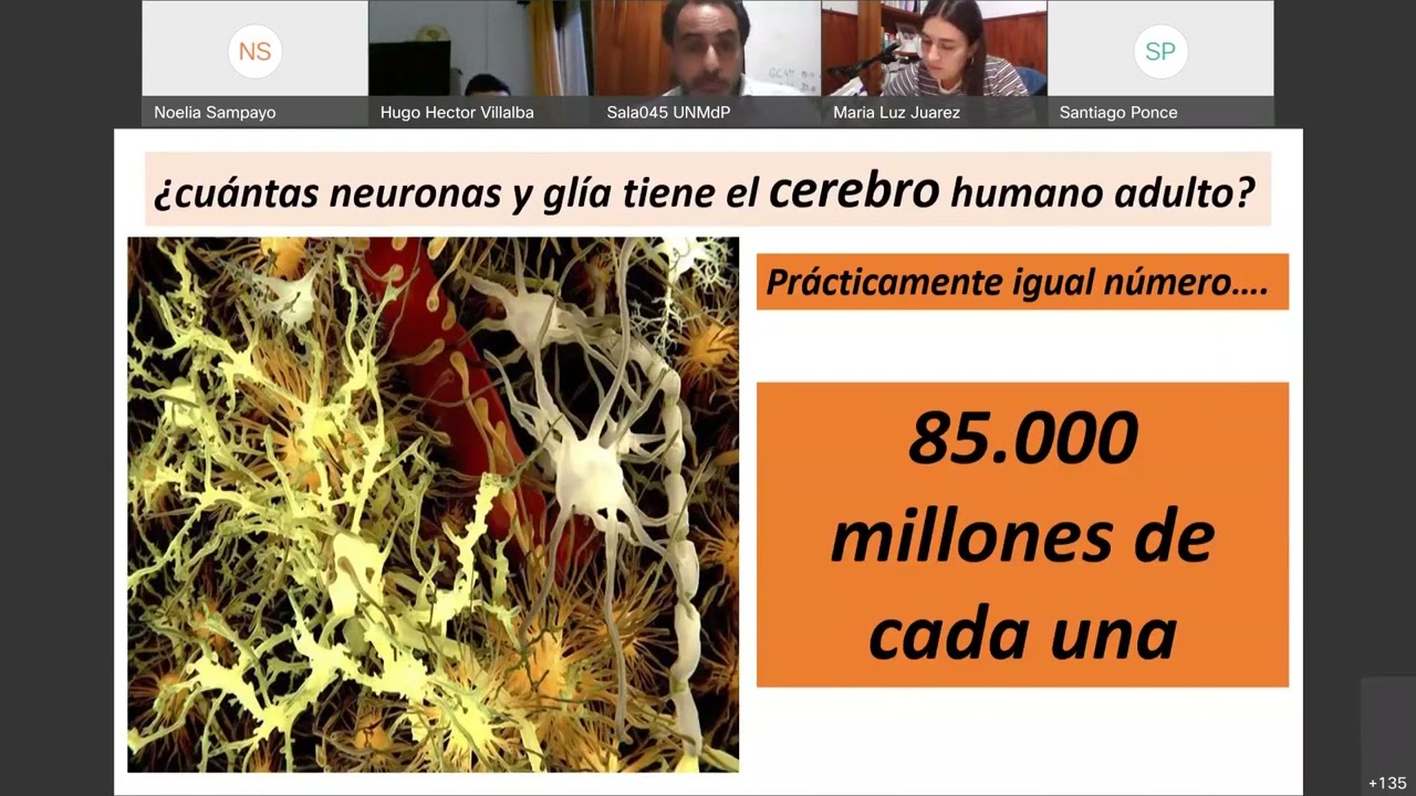
- The human brain consists of approximately 85 billion neurons and glial cells. Evolution shows variations in brain sizes and structures among species.
- Neurons initially appear in the periventricular region of the brain's cavities, migrating towards the brain's surface to form the cortex with support from astrocytes.
- During neuronal migration, trophic factors play a crucial role in guiding neurons to their destinations on the brain's surface.
Synaptic Pruning and Cortical Layer Formation
- Neurons undergo synaptic pruning to eliminate excess synapses, enhancing neural network efficiency during development.
- Prenatal stages show an increase in neuron quantity and connections before synaptic pruning occurs around ages four to six years old.
- The human cerebral cortex comprises six layers with distinct characteristics. Early migrating neurons settle in deeper layers while later ones occupy more superficial layers.
Detailed Neuroanatomy Overview
In this section, the speaker provides an overview of neuroanatomy, discussing the structure of the brain and neurons.
Understanding Brain Structures
- The telencephalon and diencephalon are separated by the corpus callosum, with structures above it derived from the telencephalon and those below from the diencephalon.
- The brain's primitive form is smooth initially but develops folds to fit within the skull as it grows faster than the cranial bones. These folds aid in accommodating the expanding brain.
Neuronal Structure and Function
- Special staining techniques are required to observe neurons under a microscope due to their unique characteristics like myelination. Different stains reveal distinct features of neurons.
- Neurons consist of a soma containing organelles like ribosomes for enzyme production. Dendrites extend from the soma, receiving signals, while axons transmit signals to other neurons or tissues.
- Axons are covered by myelin sheaths that accelerate signal conduction. Nodes of Ranvier are gaps in myelination crucial for signal transmission speed.
Importance of Myelination
- Myelin plays a vital role in enhancing signal conduction speed along axons. Diseases causing demyelination can lead to motor or sensory impairments based on which type of neuron is affected.
- Demyelinated axons conduct signals slower than myelinated ones, impacting functions such as movement coordination or sensory perception.
Neuronal Classification
- Neurons can be classified into unipolar, bipolar, or multipolar types based on their structure and function. Each type serves specific roles within the nervous system.
- Multipolar neurons are prevalent in motor functions within the central nervous system, while other types may dominate sensory systems like vision or hearing.
Neuronal Classification and Histology
In this section, the lecturer discusses neuronal classification based on structure and location, as well as histological features of neurons and supporting cells in the nervous system.
Neuronal Classification
- Neurons can be classified based on their structure into unipolar or pseudo-monopolar neurons.
- : Unipolar or pseudo-monopolar neurons have short projections that branch out.
- Pseudo-unipolar neurons are found in ganglia accompanying the spinal cord.
- : These neurons are specifically located in ganglia near the spinal cord.
Histological Features of Neurons
- Neurons can also be classified based on axon length into Golgi type I and type II neurons.
- : Type I neurons have long projections connecting distant areas like cerebral lobes to the brainstem, while type II neurons have short connections between neighboring regions.
- Microscopic examination reveals multipolar neurons with numerous extensions.
- : Multipolar neuron structures show extensive branching under an electron microscope.
Supporting Cells in Nervous System
- Various types of glial cells support neuronal function, including astrocytes, oligodendrocytes, microglia, ependymocytes, and satellite cells.
- : Astrocytes play a crucial role in forming the blood-brain barrier to protect neural tissues from harmful substances entering through the bloodstream.
Astrocyte Functions
- Astrocytes contribute to maintaining brain homeostasis by regulating substance entry into neural tissues.
- : Functions include providing structural support, forming the blood-brain barrier, phagocytizing dead nerve cells for tissue repair (gliosis), and buffering excess potassium levels.
Oligodendrocyte Role
- Oligodendrocytes primarily produce and maintain myelin sheaths around axons in the central nervous system.
Microglia and Glial Cells Function
In this section, the discussion revolves around the functions of microglia and glial cells in the central nervous system.
Microglia Function
- Microglia are similar to our phagocytic system, acting as the brain's macrophages. They derive from cells similar to monocytes and serve as a primary defense against invading microorganisms, including viruses and tumor cells.
Microglia Structure
- Microglia line the cavities of the nervous system, such as ventricles, and also cover the spinal cord cavity, forming the ependymal canal. They participate in producing cerebrospinal fluid by lining conduits where the fluid flows.
Ependymocytes Function
- Ependymal cells aid in cerebrospinal fluid formation by having cilia with high mobility that assist in fluid movement. These cells play a crucial role in facilitating liquid flow within specific structures of the nervous system.
Oligodendrocytes vs. Schwann Cells
This part delves into oligodendrocytes' role in myelination within the central nervous system compared to Schwann cells' function in peripheral nerves.
Oligodendrocyte Function
- Oligodendrocytes are akin to Schwann cells but operate within the central nervous system, providing myelin sheaths for axons. They wrap around single axons due to their inability to ensheath multiple axons simultaneously.
Glial Cells Diversity
Actions Without Myelin
In this section, the speaker discusses the presence of actions that do not involve myelin in neuronal communication.
Actions without Myelin
- Neurons can communicate electrically over short distances without the need for myelin.
- Not all axons have myelin; there are also non-myelinated axons that serve specific functions.
Axonal Pathologies and Diseases
The conversation shifts towards discussing axonal pathologies and diseases, highlighting their impact on nerve function.
Axonal Functions and Pathologies
- Axons with specific functions may be slower but serve essential purposes beyond speed.
- Pathologies affecting axons can lead to demyelination or direct destruction of the axon itself.
Multiple Sclerosis and Axonal Damage
The focus is on multiple sclerosis as a prototypical demyelinating disease and its implications for nerve health.
Multiple Sclerosis Insights
- Multiple sclerosis involves an autoimmune attack on myelin, leading to damage across various nervous system sectors.
- Considerations extend beyond demyelination to include conditions like diabetes impacting nerve health.
Vitamin B12 Deficiency and Nerve Health
The discussion delves into vitamin B12 deficiency's effects on nerve health and potential treatments.
Vitamin B12 Deficiency Implications
- Vitamin B12 deficiency can result in sensory symptoms and dementia, emphasizing its critical role in nerve health.
- Patients with diabetes experiencing nerve damage due to metabolic disorders highlight the importance of addressing nutritional deficiencies promptly.
Treatment Approaches for Nerve Pain
Exploring treatment modalities for nerve pain, including considerations around vitamin B12 supplementation.
Treatment Strategies
- While some patients report faster improvement with vitamin B12 supplementation alongside anti-inflammatory medications, scientific evidence supporting this combination remains inconclusive.
- Differentiating between neuropathic pain causes like compression versus demyelination guides appropriate treatment approaches.
Role of Vitamin B12 in Neuropathy
Addressing misconceptions around vitamin B12's role in neuropathy management and its relevance in specific conditions.
Clarifying Misconceptions
- Vitamin B12 supplementation primarily targets neuropathies caused by deficiencies rather than general compression-related issues like temporary numbness.
- Scientific validation regarding vitamin B12's efficacy in treating lumbosacral pain remains limited despite anecdotal patient experiences suggesting benefits.
Anatomy of the Brain
In this section, the speaker discusses the anatomy of the brain, focusing on structures such as the cerebellum, brainstem, ventricles, and gray and white matter.
Cerebellum and Brainstem
- The cerebellum is located posteriorly beneath the brain and is in contact with the occipital lobe. There is a structure called the tentorium that separates it from the brain.
- The brainstem is situated in front of the cerebellum and consists of structures like the pons and medulla oblongata.
Ventricle Structure
- The ventricles are cavities within the cerebral hemispheres that contain cerebrospinal fluid.
- These ventricles are visible in MRI images as black areas below the corpus callosum.
- Gray matter forms clusters known as cortical regions or cortex, while white matter comprises axons with myelin sheaths.
- Gray matter consists of neuronal cell bodies, while white matter primarily contains axons covered in myelin.
Gray and White Matter Differentiation
- Gray matter appears gray due to neuronal cell bodies present, whereas white matter looks white because of myelinated axons.
- In the brain, gray matter forms two main structures: cortex (outer layer) and nuclei (inner islands).
- Nuclei are collections of neuronal cell bodies found within gray matter regions inside the central nervous system.
- This contrasts with ganglia found outside the central nervous system in peripheral nerves.
Nerve Structure
This part delves into nerve structure detailing components like endoneurium, perineurium, epineurium, fascicles, and their roles in supporting nerve function.
Nerve Components
- Each nerve comprises multiple axons bundled together supported by connective tissue sheaths like endoneurium surrounding individual axons.
- These axon groups form fascicles enclosed by perineurium which further combine to create nerves enveloped by epineurium for structural integrity.
- Understanding these components aids in recognizing that a nerve isn't merely an individual axon but a complex assembly involving numerous axons organized within protective layers for optimal functioning.
New Section
In this section, the speaker discusses the different types of connective tissues surrounding nerve fibers.
Endoneurium, Perineurium, and Epineurium
- The endoneurium surrounds individual nerve fibers.
- Perineurium encases each fascicle.
- Epineurium encompasses the entire peripheral nerve.
New Section
This part focuses on a macroscopic cross-section of the spinal cord, highlighting the arrangement of gray and white matter.
Gray and White Matter in Spinal Cord
- Gray matter is centrally located and resembles a butterfly shape.
- White matter surrounds gray matter entirely.
- Gray matter contains neuronal cell bodies while white matter consists of axons for transmitting signals.
New Section
The discussion delves into the composition and functions of gray matter within the spinal cord.
Composition of Spinal Cord
- White matter facilitates axonal transmission up and down.
- Gray matter houses neuronal cell bodies with varied functions.
- Gray matter forms horns: ventral (anterior), lateral, and dorsal (posterior).
New Section
Here, the speaker elaborates on the structure of gray matter within the spinal cord.
Structure of Gray Matter
- Gray matter resembles a butterfly shape with a central ependymal canal.
- Divided into ventral (anterior), lateral, and dorsal (posterior) horns.
- Posterior horn primarily processes sensory information while anterior horn deals with motor neurons.
New Section
This segment explores how gray and white matters are organized within the spinal cord for specific functions.
Functions in Gray Matter
- Posterior horn relates to sensory input processing.
- Anterior horn involves motor neurons for muscle innervation.
- White matter divides into dorsal, lateral, and ventral columns for distinct actions.
New Section
The speaker explains how different types of actions are carried out by various components within the spinal cord's gray and white matters.
Actions in Spinal Cord
- Different functions occur in anterior (ventral) vs. posterior (dorsal) regions based on motor or sensory roles.
Concepts of the Nervous System
In this section, the speaker discusses the concepts related to the nervous system, focusing on sensory and motor functions, as well as the classification of the central and peripheral nervous systems.
Understanding Sensory and Motor Functions
- Sensory functions involve information entering the nervous system.
- Sensory inputs are sensitive actions that bring information into the nervous system.
- Motor functions relate to actions exiting the nervous system.
- Motor actions carry information outwards from the nervous system.
Classification of Nervous System Components
- The nervous system is divided into two main parts: central and peripheral.
- The central nervous system comprises the brain and spinal cord.
- The peripheral nervous system consists of nerves, ganglia, and plexuses.
Clarification on Nervous System Components
- Differentiating between central and peripheral components can be challenging.
- Understanding structures like plexuses and nerve roots aids in classification.
- The location of neuronal cell bodies determines central or peripheral affiliation.
- Neuronal cell bodies within nerves are part of the central nervous system.
Neuronal Bodies in Central vs. Peripheral Systems
This segment delves into the distribution of neuronal bodies within the central and peripheral nervous systems, emphasizing their distinct roles.
Location of Neuronal Bodies
- Neuronal bodies primarily reside in the central nervous system.
- Central neurons' bodies form a significant part of this neural network.
Role Distribution in Central vs. Peripheral Systems
- Axons extend from neuronal bodies for signal transmission.
- Axons are essential for transmitting signals within neural networks.
- Distinctions exist between central and peripheral systems based on body locations.
- The central nervous system is encased by bony structures for protection.
Impact of Diseases on Nervous System Components
This section explores how diseases affect different components of the nervous system, highlighting variations in damage patterns based on disease types.
Disease Effects on Neural Structures
- Mylelinating diseases impact specific neural components differently.
- Diseases may target either central or peripheral neural structures uniquely.
Vitamin Deficiency Consequences
- Vitamin B12 deficiency can lead to diverse neurological symptoms due to axon demyelination.
New Section
In this section, the discussion revolves around the formation of neuronal structures in the central and peripheral nervous systems.
Neuronal Structures Formation
- The cortex or core forms a nucleus in the central nervous system, while neuronal bodies in the peripheral nervous system form ganglia.
- Neuronal bodies in the peripheral nervous system are either located in ganglia or within the central nervous system.
New Section
This part delves into the relationship between axons and neuronal bodies in the peripheral nervous system.
Axons and Neuronal Bodies
- Axons are not solitary but have associated neuronal bodies, which are found either in ganglia (peripheral nervous system) or within the central nervous system.
- The gray matter is formed when multiple neuronal bodies gather, representing a change from white matter to gray matter.
New Section
Exploring how cortical structures relate to actions transmitted by neurons.
Cortical Structures and Neuronal Actions
- The cortex of the brain and brainstem play a role in sending out actions initiated by neuronal bodies present there.
Understanding the Central Nervous System
In this section, the speaker delves into the components of the central nervous system, focusing on the spinal cord and peripheral nervous system.
Components of the Central Nervous System
- The spinal canal contains meninges that encase the spinal cord, including the structure known as "cola de caballo," which comprises roots emanating from the spinal cord within the vertebral canal.
- Different types of nerve fibers exist within the central nervous system, such as association fibers connecting regions within a hemisphere, commissural fibers linking hemispheres like the corpus callosum, and projection fibers extending to distant areas like the brainstem or spinal cord.
Organization of Axonal Fibers in Spinal Cord
- Within the spinal cord, white matter surrounds gray matter forming a butterfly shape. Bundles of axons grouped together are termed fascicles when they share a common origin, trajectory, and destination.
- Fascicles consist of axons originating from various locations but converging during their course before diverging again towards different destinations. This arrangement allows for distinct origins and destinations while sharing a common pathway.
Exploring Brain Structure: External Configuration
The discussion shifts to exploring external features of the brain, particularly focusing on its surface configuration.
External Features of Brain
- The two cerebral hemispheres are separated by a cerebral fissure that cannot be easily divided due to commissural fibers like the corpus callosum connecting them.
New Section
In this section, the speaker discusses the structures of the brain visible through a cerebral fissure, highlighting key components such as the brainstem, optic nerve, optic chiasm, hypothalamus, and pituitary gland.
Structures Visible in the Cerebral Fissure
- The cerebral fissure reveals the brainstem passing through it.
- Components visible include the eyeballs, optic nerve marked as number 3 5, optic chiasm, hypothalamus (cut for removal), and pituitary gland left inside.
New Section
This part focuses on the external face of the cerebral hemisphere and emphasizes understanding fissures that divide lobes and sulci that further divide lobes into gyri.
External Face of Cerebral Hemisphere
- Learning about fissures that divide hemispheres into lobes.
- Fissures are deep divisions while sulci are shallower grooves within lobes dividing them into gyri.
New Section
Exploring how fissures on the external face delineate lobes and sulci within lobes define gyri in detail.
Fissures and Sulci on External Face
- Fissures on external face delimit lobes while sulci within lobes define gyri.
- Understanding how these structural features contribute to brain organization.
New Section
Continuing to examine fissures on the external face of the brain to understand their role in defining lobes and gyri.
Significance of Fissures and Gyri
- External face exhibits fissures demarcating lobes and sulci defining gyri.
- Observing three main fissures - lateral fissure (Silvian), central fissure (Roland), parietooccipital or transverse occipital fissure externally.
New Section
Detailed exploration of specific cerebral fissures including lateral (Silvian) fissure's origin from inferior brain surface to its appearance on external surface.
Understanding Lateral Fissure
- Lateral or Silvian fissure originates from inferior brain surface.
- It houses an important artery called middle cerebral artery known as Sylvian artery too.
New Section
In this section, the speaker discusses the different lobes of the brain and their locations.
Lobes of the Brain
- The occipital lobe is located behind the parietal lobe. The external perpendicular runs through these lobes.
- The frontal lobe is above and in front, while the temporal lobe is below the parietal lobe.
- There is a hidden lobe called the insular lobe within the Sylvian fissure.
- Opening the Sylvian fissure reveals the insular lobe, resembling an island of brain tissue.
New Section
This part focuses on the frontal lobe's structure and its divisions.
Frontal Lobe Structure
- The frontal lobe has three main sulci: superior frontal sulcus, inferior frontal sulcus, and ascending frontal sulcus.
- These sulci divide the frontal lobe into superior, middle, and inferior gyri.
- The gyri are named based on their position relative to these sulci for anatomical reference.
New Section
Exploring specific areas within brain structures related to language processing.
Language Processing Areas
- Within the inferior frontal gyrus lies Broca's area responsible for speech expression.
- Damage to Broca's area can result in expressive aphasia where speech expression is impaired.
Area Motora Primaria
In this section, the speaker explains the primary motor area of the brain and its role in voluntary movement control.
Primary Motor Area Function
- The primary motor area is named as such because it contains the cell bodies of neurons responsible for voluntary movement.
- Located in the left hemisphere, this area controls voluntary movements of the right side of the body from the waist up.
- Specifically, within the left hemisphere's primary motor area lies the frontal ascending convolution, regulating movements on the external side of the brain from waist up.
Motor Areas and Decision Making
This part delves into additional motor areas beyond the primary motor area and discusses regions involved in decision-making processes.
Motor Supplementary Areas
- Apart from the primary motor area, there are supplementary and premotor areas responsible for motor planning.
- These areas also contribute to cognitive functions and decision-making processes within the brain.
Broca's Area and Language Expression
The discussion shifts towards Broca's area, its significance in language expression, and potential implications of lesions in this region.
Broca's Area Functionality
- Broca's area, primarily located in the left hemisphere, governs speech expression. Lesions here can lead to expressive language loss known as Broca's aphasia.
- Contrarily, damage to Broca's area in the right hemisphere results in a condition called aprosodia characterized by intonation deficits rather than speech expression impairment.
Importance of Intonation
Emphasizing intonation importance in human communication compared to animals.
Significance of Intonation
- Intonation plays a crucial role in human communication nuances; it resides predominantly in the right hemisphere’s frontal inferior convolution.
New Section
In this section, the discussion revolves around the loss of mobility in the upper limbs due to cultural factors affecting one side of the body and its implications on muscle function.
Cultural Factors and Mobility Loss
- The loss of mobility in the upper limb on one side can affect not only that limb but also all muscles from the waist upwards.
New Section
This part delves into the complexities of muscle movement in relation to brain lesions and voluntary actions.
Muscle Movement and Brain Lesions
- Difficulty arises when trying to move one set of paravertebral muscles while keeping another set still due to brain lesions.
New Section
The conversation shifts towards brain hemispheres, specifically focusing on Broca's area and its impact on speech expression.
Broca's Area and Speech Expression
- Lesions in Broca's area can lead to speech expression issues, with different effects depending on which hemisphere is affected.
New Section
This segment explores how Broca's area localization differs between right-handed and left-handed individuals.
Localization of Broca's Area
- While most right-handed individuals have Broca's area in the left hemisphere, about a quarter of left-handed individuals have it in the right hemisphere.
New Section
The discussion continues by emphasizing the predominant localization of Broca's area in specific brain hemispheres.
Predominant Localization of Broca's Area
- The majority of people have their Broca's area located in the left hemisphere, regardless of handedness.
New Section
Moving on to explore the temporal lobe structure, particularly focusing on its divisions based on sulci and gyri.
Structure of Temporal Lobe
- The temporal lobe is divided into superior and inferior temporal sulci, further segmented into three gyri: superior, middle, and inferior temporal gyri.
New Section
This part elaborates on specific areas within the superior temporal gyrus related to auditory functions.
Auditory Functions in Superior Temporal Gyrus
- The primary auditory area resides within the superior temporal gyrus, responsible for processing incoming auditory stimuli.
New Section
Further details are provided regarding how auditory information is processed across brain hemispheres within specific regions.
Auditory Information Processing
- Auditory information from both ears reaches distinct regions within each hemisphere for processing, highlighting specialized functions based on input sources.
New Section
Discussion continues regarding auditory processing areas' resilience to unilateral damage within specific brain regions.
Resilience to Unilateral Damage
- Damage to one side’s superior temporal gyrus does not result in deafness due to bilateral auditory information reception by corresponding regions in both hemispheres.
New Section
Exploring potential outcomes following damage or impairment within critical auditory processing areas.
Outcomes Following Impairment
Understanding the Occipital and Parietal Lobes
In this section, the speaker delves into the anatomy of the occipital and parietal lobes, discussing specific sulci, gyri, and their functions.
Occipital Lobe Anatomy
- The occipital lobe contains two main sulci: superior occipital sulcus and occipital sulcus. These sulci delineate three convolutions: superior, middle, and inferior occipital gyri.
- The external face of the occipital lobe houses the visual recognition area responsible for identifying objects visually. This area aids in recognizing objects like houses, clocks, or cars.
- Lesions in the occipital lobe's external face can lead to visual agnosia, causing a lack of recognition of visual stimuli despite intact vision.
Visual Memory and Recognition
- Visual memory is located on the external face of the occipital lobe alongside visual recognition areas. Lesions in this region can result in deficits in visual memory and recognition abilities.
- Damage to specific areas within the occipital lobe can lead to selective impairments such as agnosia without affecting overall vision.
Exploring Parietal Lobe Functions
The discussion shifts towards detailing the functions and structures within the parietal lobe.
Parietal Lobe Structure
- The parietal lobe features a single prominent sulcus known as intraparietal or interparietal sulcus that divides it into distinct regions.
- This sulcus gives rise to a convolution called parietal ascending convolution which plays a role in primary sensory processing. It is situated between Rolando's fissure anteriorly and parietoascendant fissure posteriorly.
Sensory Processing in Parietal Lobe
- The parietal ascending convolution serves as the primary sensory area where sensory information from touch or heat reaches contralaterally (opposite side). This region processes sensations from waist upwards only.
Understanding Brain Lobes and Functions
In this section, the speaker discusses the different brain lobes and their functions, focusing on areas such as the frontal lobe, temporal lobe, parietal lobe, and occipital lobe.
Frontal Lobe Functions
- The frontal lobe includes the superior and inferior frontal sulci.
- These sulci delineate the superior and inferior frontal gyri.
- Areas within the frontal lobe are crucial for specific functions:
- Broca's area in the left hemisphere is responsible for speech expression.
- The precentral gyrus houses the primary motor area controlling movements from waist upwards contralaterally.
Temporal Lobe Functions
- The temporal lobe contains essential regions:
- Wernicke's area for language comprehension.
- The superior temporal gyrus hosts the primary auditory area.
Parietal Lobe Functions
- Within the parietal lobe:
- The postcentral gyrus holds the primary sensory area receiving information on touch, temperature, pain, and body position (proprioception).
Occipital Lobe Function
- In the occipital lobe:
- Recognition of visual stimuli occurs primarily on its external surface.
Motor Areas in Brain Lobes
This segment delves into motor areas within brain lobes, emphasizing the importance of understanding cortical organization for movement coordination.
Motor Areas in Brain Lobes
- The primary motor area is located in the frontal ascending gyrus.
- Other motor areas exist beyond just this primary region.
- Neurons in these areas organize to execute voluntary movements efficiently.
- These neurons' bodies are situated superficially in cortical layers.
Brain Hemisphere Communication
Here, communication between brain hemispheres regarding auditory processing is explored along with insights into hearing impairments.
Auditory Processing Across Hemispheres
- Hearing impairment typically stems from issues in inner ear structures or auditory nerve rather than cerebral lesions.
- Both hemispheres receive auditory input bilaterally to prevent complete deafness even if one side is affected.
Fasciculus Architecture and Function
This part elucidates fasciculus structure connecting key brain areas like Broca's and Wernicke's regions.
Fasciculus Composition and Role
- The arcuate fasciculus links Broca's area with Wernicke's region facilitating language processing.
Understanding Brain Functionality
In this section, the speaker delves into the connectivity within the brain, focusing on areas like Broca's and Wernicke's areas and how they are associated with language processing.
The Fasciculus
- The fascicle 4 comprises association fibers linking two areas within the same hemisphere.
- It connects regions such as Broca's area and Wernicke's area, facilitating language functions.
- Neurons in Wernicke's area send axons towards Broca's area for synaptic connections.
Functional Studies
- Functional MRI and tractography enable detailed studies of brain connectivity.
- These techniques aid in identifying specific brain areas responsible for functions like reading comprehension.
Neural Plasticity
- The brain adapts through neural plasticity, forming new connections throughout life.
- Learning a new skill or language involves creating new neural networks regardless of age.
Neural Networks and Gender Differences
This segment explores neural network formation, gender-based brain differences, and myths surrounding neuronal capabilities.
Importance of Neural Connections
- Having numerous neurons is insufficient without well-established neural networks.
- Effective neural connections are crucial for cognitive abilities rather than neuron quantity alone.
Gender Variances in Brain Connectivity
- Women exhibit superior inter-hemispheric and intra-hemispheric connectivity compared to men.
- Enhanced connectivity aids women in integrating concepts efficiently across brain regions.
Myths Debunked
- Contrary to popular belief, women possess robust neural networks that enhance cognitive processes.
Neurophysiology Insights
In this section, the speaker delves into neurophysiology concepts related to neural plasticity and sensory systems.
Neural Plasticity and Sensory Systems
- Neurons in the brain can reorient themselves when a body part is amputated, with neurons previously dedicated to the missing limb adapting to serve other functions, such as facial sensations.
- Neurons are dynamic entities that can establish numerous connections with other neurons, highlighting the brain's capacity for intricate neural networks.
- Discussion on phantom limb sensation post-amputation, attributing it to cortical reorganization where neurons from the amputated area connect with neighboring neurons, leading to sensory illusions.
- The phenomenon of phantom limb sensation is explained by the brain's adaptation post-amputation, where neurons repurpose their function and create new connections in adjacent areas.
- Phantom limb experiences typically diminish over time due to neural reorientation processes; emphasizing that such sensations are not lifelong but rather a result of adaptive neural mechanisms.
The Brain's Role Beyond Neurocentrism
This segment explores the speaker's perspective on neurocentrism and emphasizes the broader influences on brain development beyond traditional neuroscience views.
Beyond Neurocentrism
- Critique of neurocentrism portraying the brain as a deity-like entity; instead, highlighting that our interactions and experiences shape our brain through forming neural networks based on stimuli like touch and social interactions.
- Emphasis on how sensory experiences drive brain development rather than the brain governing sensations; distinguishing between neurophysiology focusing on bodily responses versus neurocentrism centered solely on cerebral functions.