Knee MRI scan protocols, positioning and planning
New Section
This section provides instructions for conducting a knee MRI scan, including safety checks, patient positioning, and protocol selection.
Conducting the Scan
- Before bringing the patient into the scanner room, perform all necessary safety checks.
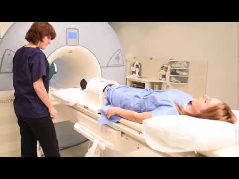
- Help the patient lie supine on the scanner table with the effective knee in the dedicated knee coil. The apex of the patella should be about a centimeter below the center of the coil.
- Assemble the coil and place pads to ensure patient comfort.
- Provide the patient with an emergency buzzer and ear protection to reduce scanner noise.
- Move the table and center the laser beam localizer over the lower border of the patella.
- Ensure that the patient is calm and comfortable before leaving them in the scanner room.
Patient Details and Protocol Selection
- In the control room, select or manually enter correct patient details in your browser. Accuracy is crucial for SAR calculation.
- Register that patient is lying feet first and supine.
- Choose an appropriate knee protocol based on hospital and radiologist guidelines.
- Begin with an axial localizer sequence from ACA localizer. Plan sagittal and coronal localizers parallel and perpendicular to medial and lateral condyles.
Diagnostic Sequences
Proton Density (PD) Fat Saturated Axial Sequence
- Angle positioning box perpendicular to long axis of femur and tibia on sagittal localizer.
- Ensure block covers area from above patella to tibial tuberosity.
- On coronal view, angle slices parallel to tibial plateau using saturation bands above and below axial block.
Sagittal Views
- First sagittal sequence is STIR (Short Tau Inversion Recovery) designed to suppress fat signal on HCL plane.
- Plan slices parallel to lateral condyle of femur, which is usually parallel to the ACL (anterior cruciate ligament).
- On coronal view, align slices parallel to midline of femur and tibia.
Reviewing Images
- Start reviewing images once planning is completed.
- PD Fat Saturated Axial Image: Fluids appear bright, while fat appears dark. Useful for visualizing cartilage, ligaments, and bony edema.
- PD Fat Saturated Coronal Image: Provides excellent visualization of meniscal tears and ligaments.
- Sagittal Images: Allow visualization of menisci, ACL, PCL, tears, and insufficiencies.
- T1 Sagittal Image: Useful in diagnosing cystic and blood components.
- T2 Star or T2 Gradient Echo Sagittal Image: Useful for visualizing menisci and assessing ACL and PCL.
The summary has been provided in English as requested.