✅ EMBRIOLOGÍA del SISTEMA NERVIOSO CENTRAL (Parte 1°) 🧠⚡
Embryology of the Central Nervous System
Introduction to Neural Development
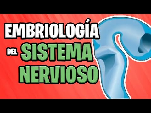
- The central nervous system (CNS) begins to form in the third week of embryonic development as a thickened ectoderm plate known as the Neural Plate.
- This plate is located in the mid-dorsal region, anterior to the primitive ganglion, and its lateral edges rise to create the Neural Folds.
Formation of the Neural Tube
- Fusion of the Neural Folds starts in the cervical region and progresses both cranially and caudally, forming open ends called neuropores that connect with the amniotic cavity.
- The cranial neuropore closes first, beginning from two sites: one at cervical closure and another emerging later in the forebrain.
Key Milestones in Closure
- Definitive closure of the cranial neuropore occurs around day 25 (18-20 somites), while closure of the caudal neuropore happens three days later.
- The cephalic end of the neural tube develops into three primary brain vesicles: forebrain, midbrain, and hindbrain.
Differentiation into Secondary Vesicles
- By week five, these primary vesicles differentiate into five secondary vesicles:
- Forebrain → telencephalon & diencephalon
- Midbrain remains unchanged
- Hindbrain → metencephalon & myelencephalon
Brain Structure Development
- Major structures derived from these vesicles include:
- Telencephalon: Cerebral hemispheres
- Diencephalon: Optic vesicle, thalamus, hypothalamus, pituitary gland
- Midbrain: Anterior and posterior colliculi
- Metencephalon: Cerebellum & pons (bridge)
- Myelencephalon: Medulla oblongata
Ventricular System Formation
- The spinal cord's central canal connects with brain cavities:
- Hindbrain forms fourth ventricle,
- Diencephalon forms third ventricle,
- Cerebral hemispheres develop lateral ventricles.