PLEXO LOMBAR
Understanding the Lumbosacral Plexus
Overview of the Lumbosacral Plexus
- The lumbosacral plexus is often referred to as a combined structure, encompassing both lumbar and sacral components, indicating its continuity in nerve fibers.
- The coccygeal plexus is notably smaller, consisting mainly of one nerve component; its functions will be discussed later.
Nerve Origins and Functions
- Peripheral nerves originating from the lumbosacral plexus are responsible for somatic and sensory innervation in the lower body.
- The lumbosacral plexus includes roots from L1 to S4, with some authors also mentioning AS5, highlighting variations in scholarly perspectives.
Anatomy and Innervation
- The coccygeal plexus arises from S4-S5 roots, contributing to a pair of nerves that serve the coccyx area.
- Nerves from the sacral segment innervate the perineum, which houses both urogenital and anal regions.
Clinical Examination Techniques
- To assess integrity within the lumbosacral plexus during clinical examinations, practitioners evaluate motor functions through muscle actions and sensory responses via dermatomes.
- Symptoms such as pain or altered sensations can indicate issues related to spinal nerves or conditions like herniated discs affecting lower limb function.
Identifying Sensory Regions
- Recognizing specific dermatomes is crucial for diagnosing nerve-related symptoms in lower limbs; overlapping innervation must be considered for accurate assessments.
Dermatome and Myotome Overview
Sensory Reception and Dermatomes
- The discussion begins with the importance of general sensitivity, particularly thermal sensitivity (heat and cold), touch, and pressure. These are essential for testing sensory integrity through dermatomes.
- The inguinal ligament is highlighted as a key anatomical landmark related to the L1 dermatome, aiding in clinical assessments.
- Anatomical markers such as the greater trochanter are used to define regions of innervation for various dermatomes, including L2 on the lateral thigh.
- The medial aspect of the first toe is associated with the L3 dermatome, while L4 covers areas like the inner knee and medial malleolus.
- The territory of innervation extends to include interdigital regions between toes, specifically noting that L5 affects areas between the first and second toes.
Posterior Regions and Reflex Testing
- The fifth toe's lateral aspect corresponds to S1 dermatome territory; this section also discusses posterior leg regions relevant to S2.
- A focus on gluteal regions indicates that T3i relates to specific skin territories overlying these areas.
- Key dermatomes S4 and S5 are tested in perineal areas, which include urogenital structures crucial for understanding sensory function in these zones.
- Discussion includes reflex testing methods like cutaneous anal reflex using cotton swabs around perianal muscles to assess normal function.
Understanding Innervation Patterns
- Knowledge of anatomical territories helps localize potential issues within nerve roots by mapping out affected dermatomes based on symptoms presented.
- An emphasis is placed on understanding how each nerve root contributes to specific muscle groups' innervation in lower limbs, enhancing diagnostic capabilities.
Myotomes: Muscle Innervation Insights
- Reference is made to figures illustrating myotomes responsible for muscle innervation in lower limbs; these visuals aid comprehension of complex relationships between nerves and movements.
- Detailed descriptions outline how different movements at joints correspond with specific nerve roots (e.g., flexion/extension at hip joint linked with L2-L5).
Movement Mechanics Related to Nerve Roots
- Each movement at joints (like hip flexion or knee extension) is governed by particular nerve roots; this relationship underscores functional anatomy's relevance in clinical practice.
- Specific movements such as abduction or internal/external rotation at hip joints are tied back to their respective nerve root contributions (L3-L5).
Joint Movements: Flexion and Extension Dynamics
- Knee joint mechanics are discussed concerning extensions dominated by L3-L4 while flexions involve L5-S1 interactions during activities like driving or walking.
- Dorsiflexion versus plantarflexion dynamics highlight how various actions engage different nerve roots across foot movements.
Understanding the Lumbar Plexus and Nerve Roots
Overview of Nerve Roots and Movements
- The discussion begins with an overview of nerve roots, specifically focusing on L1 to S2, highlighting their roles in flexion and extension movements.
- Emphasis is placed on testing the integrity of nerve roots during knee flexion and extension, particularly involving L1 and L2 for hip flexion.
- Dorsiflexion and plantarflexion are discussed, noting that S1 and S2 are primarily responsible for these movements.
Testing Integrity of the Nervous System
- The importance of understanding L5 root involvement is highlighted, especially concerning the fibular nerve's role in motor function.
- The intrinsic muscles of the foot are examined; S2 and S3 are identified as key contributors to toe movements.
- A comprehensive approach to testing lumbar-sacral plexus integrity through dermatomes is introduced.
Reflex Testing Techniques
- Methods for assessing reflexes include percussion tests on the Achilles tendon (S1/S2) and patellar tendon (L3/L4).
- Observations from these tests can indicate potential issues with specific nerve roots based on muscle responses.
Pathology Insights: Herniated Discs
- Discussion shifts to herniated discs affecting lumbar regions, detailing how they compress surrounding structures leading to symptoms like paresthesia or motor function loss.
- Visual aids illustrate disc herniation impacts on spinal nerves, emphasizing symptomatology related to decreased reflexes and muscle tone.
Degenerative Changes in Spinal Anatomy
- An exploration into degenerative changes within intervertebral foramina highlights stenosis effects on nerve root compression.
- Tumor-like growth patterns may also contribute to similar compressive symptoms observed in patients.
Exploring the Lumbar Plexus Structure
Components of the Lumbar Plexus
- The foundational question regarding which roots form the lumbar plexus is addressed: L1 through L4 are confirmed as primary contributors.
- Contributions from T12 are acknowledged; anatomical relationships between abdominal structures and lower limb innervation are outlined.
Anatomical Organization
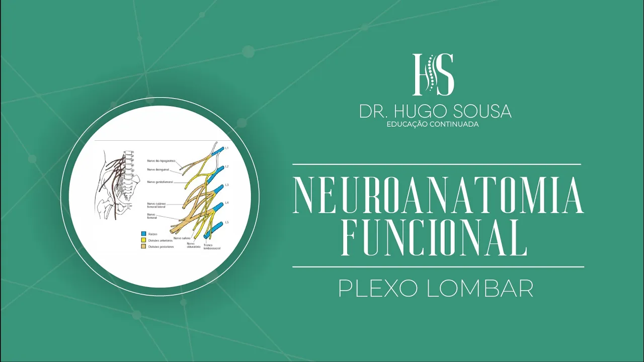
- Detailed descriptions clarify divisions within the plexus—posterior (yellow representation), anterior (green representation)—and their functional implications.
Formation of Key Nerves
- Specific nerves such as genitofemoral arise from combinations of L2/L3 roots; this section emphasizes anatomical connections crucial for understanding lower limb innervation.
Nerve Anatomy and Function
Overview of Muscle and Nerve Roots
- Discussion on the thick structure of muscle roots, highlighting various muscular branches that emerge from them.
- Introduction to the obturator nerve, detailing its origin at the lumbar level (L2, L3, L4) and its primary function in adduction of the thigh.
Complexity of the Lumbar Plexus
- Comparison between lumbar plexus complexity versus brachial plexus; emphasis on simpler structure in lumbar region.
- Explanation of posterior and anterior divisions within the lumbar plexus, noting a higher number of nerves in the posterior division.
Key Nerves and Their Functions
- Identification of key nerves such as hypogastric (T12, L1), genitofemoral (L1, L2), femoral (L2-L4), and their respective roles.
- Description of how these nerves traverse through anatomical structures like the obturator canal.
Anatomical Relationships
- Visualization of nerve locations relative to pelvic wall muscles including quadratus lumborum and iliacus.
- Discussion on how lumbar plexus roots are positioned posteriorly to certain muscles like psoas major.
Medial Nerve Pathways
- Introduction to the lumbosacral trunk formed by roots L4-L5; discussion on its significance in nerve pathways.
- Examination of medial relationships among various nerves including iliohypogastric and ilioinguinal.
Functions of Specific Nerves
Iliohypogastric Nerve
- Overview of iliohypogastric nerve's location lateral to psoas major; involvement with abdominal wall muscles.
- In-depth look at its innervation territory affecting lower abdominal skin and associated muscle groups.
Ilioinguinal Nerve
- Description of ilioinguinal nerve's segmental origin (L1); details about its sensory innervation over medial thigh area and scrotum.
Genitofemoral Nerve
Nerve Innervation and Reflexes in the Lower Body
Overview of Nerve Functions
- The discussion begins with the innervation of the scrotum, highlighting its relation to the anterior thigh skin and labia majora in women. The cremaster muscle is mentioned for its role in elevating the testicles.
Testing Reflexes
- A demonstration of the cremasteric reflex is introduced, which can be tested by stimulating the anterior medial abdomen. This reflex involves sensory input from specific nerves.
Lateral Cutaneous Nerve of Thigh
- The lateral cutaneous nerve (L2-L3) innervates the anterior and lateral surfaces of the thigh. Its pathway is described as passing below the inguinal ligament.
Meralgia Paresthetica
- Conditions such as meralgia paresthetica are discussed, often caused by tight clothing or prolonged sitting, leading to symptoms like paresthesia due to nerve compression.
Sensory and Motor Functions of Femoral Nerve
- The femoral nerve's extensive sensory territory includes parts of the leg and foot. It also innervates key muscles such as quadriceps femoris (vastus medialis, vastus lateralis, vastus intermedius, rectus femoris).
Additional Muscles Innervated by Femoral Nerve
- Besides quadriceps, other muscles like iliopsoas are also innervated by this nerve. The sartorius muscle is highlighted for its significant length and flexion action at the hip joint.
Obturator Nerve Functionality
- The obturator nerve (L2-L4 roots) provides sensation to the medial thigh surface and motor function to adductor muscles including adductor longus, brevis, magnus, and obturator externus.
Accessory Obturator Nerve Insights
- When present, an accessory obturator nerve crosses under inguinal ligament near psoas major. Its branches serve various functions related to knee articulation sensitivity.
Summary of Key Nervous Structures
- An overview concludes with a recap on cutaneous nerves: lateral femoral cutaneous nerve contributes sensory information from anterior-medial thigh; saphenous nerve extends sensory supply to inner knee area down through medial leg and foot regions.
Plexus and Nerve Innervation
Overview of Sacral Plexus and Gluteal Region Innervation
- The discussion begins with the relationship between the sacral plexus and gluteal region, focusing on nerves that innervate this area.
- It is clarified that the cluneal nerves are involved in the innervation of the superior and lateral aspects of the gluteal region, originating from lumbar roots L1, L2, and L3.
- The middle cluneal nerves derive from posterior roots S1, S2, and S3 through the posterior sacral foramina, indicating their role in sacral plexus formation.
- Inferior cluneal nerves also play a significant role in innervating lower regions of the gluteal area; they originate from cutaneous nerve branches.