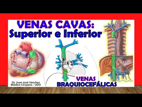
🥇 VENA CAVA SUPERIOR e INFERIOR, Anatomía. Venas BRAQUIOCEFÁLICAS, Fácil y Sencillo.
Understanding the Venous System: The Superior and Inferior Vena Cava
Overview of the Venous System
- Introduction to the topic of major thoracic vessels, focusing on the venous system, specifically the superior and inferior vena cava.
- Mention of brachiocephalic veins as essential components related to the superior vena cava.
Brachiocephalic Veins
- Explanation of brachiocephalic veins, previously known as innominate veins, formed by the union of internal jugular and subclavian veins.
- Description of anatomical differences between right and left brachiocephalic veins; right is shorter due to its position relative to the heart.
Formation and Drainage
- Both brachiocephalic veins converge behind the first costal cartilage on their respective sides to form the superior vena cava.
- The right brachiocephalic vein drains blood from both head/neck (via internal jugular vein) and upper limb (via subclavian vein).
Anatomical Relationships
- Discussion on how anatomical positioning affects catheter placement in medical procedures; easier access via right side due to vertical orientation.
- Left brachiocephalic vein has a more horizontal or oblique course compared to its right counterpart.
Clinical Relevance
- Importance of understanding anatomical relationships for clinical practices such as catheterization; left passes anteriorly over branches from aorta.
Understanding the Venous Drainage of the Thorax
Overview of Intercostal Veins
- The left superior intercostal vein drains into the left brachiocephalic vein, while the right superior intercostal vein does not follow this pattern.
- The vertebral vein also drains into both brachiocephalic veins, alongside the inferior thyroid vein.
Internal Mammary and Pericardial Veins
- Internal mammary veins specifically drain into the brachiocephalic vein; additionally, a cut section shows the pericardial veins draining into both sides' brachiocephalic veins.
Introduction to Superior Vena Cava
- Transitioning to discussing the superior vena cava, which drains a larger area than the brachiocephalic veins due to its role in collecting blood from various regions including thoracic cavity walls and viscera.
Formation and Function of Superior Vena Cava
- The superior vena cava is formed by the union of both brachiocephalic veins and begins at the level posterior to the first costal cartilage on the right side.
- It carries venous blood from designated areas directly to the right atrium of the heart.
Anatomical Relationships of Superior Vena Cava
- The superior vena cava is surrounded by parietal pericardium; some authors differentiate between an extrapercardial portion and an intrapericardial portion.
- Important anterior relations include thymus gland, sternum, and first two costal cartilages; posteriorly it relates closely with trachea and main bronchus.
Lateral Relations and Afluents
- Laterally, it is associated with phrenic nerve and pericardiophrenic vessels; pleura mediastinalis also has lateral relations with it.
Vena Cava: Anatomical Insights
Overview of the Superior and Inferior Vena Cava
- The superior vena cava is a valvular vein that lacks valves at its entry point into the heart, allowing venous return without obstruction.
- The inferior vena cava is significantly longer than the superior, collecting blood from the abdominal cavity, pelvic cavity, and lower limbs.
- The inferior vena cava originates from the union of the common iliac veins and begins at vertebra L4, indicating its anatomical starting point.
Pathway and Structure of the Inferior Vena Cava
- It drains blood from lower body regions into the right atrium of the heart after perforating the diaphragm through a specific orifice.
- This orifice in the diaphragm allows passage for blood flow while separating thoracic and abdominal cavities.
Key Dilatations Along Its Course
- There are two notable dilatations in the inferior vena cava: one where hepatic veins drain (hepatic sinus) and another where renal veins enter (renal sinus).
- The inferior vena cava enters directly into the right atrium, similar to how the superior vena cava does.
Valves and Portions of Vena Cava
- Unlike its counterpart, the inferior vena cava has a vestigial valve known as Eustachian valve located at its entrance to the right atrium; however, it serves no significant function.
- The structure consists of three portions: thoracic (superior), diaphragmatic (shorter), and abdominal (longest).
Anatomical Relationships
- The abdominal portion is retroperitoneal, meaning it lies behind peritoneal lining; both it and abdominal aorta are confined to posterior wall of abdomen.
- Its position shifts towards right side as it ascends due to drainage into right heart chambers; maintains relationships with lumbar diaphragm portions.
Surrounding Structures
- Anteriorly related structures include gonadal arteries (ovarian/testicular), while posterior relations involve adrenal glands and upper kidney poles.
Anatomical Relationships of the Liver and Surrounding Structures
Overview of Liver Positioning
- The new area of the liver is located posterior to several key structures: the pancreas, portal triad, and the third portion of the duodenum (transverse section).
- It is also positioned behind the root of the mesentery and superior mesenteric vessels (artery and vein).
Relationship with Duodenum
- The liver has a lateral right relationship with the second portion of the duodenum, specifically its descending part.
Venous Drainage into Inferior Vena Cava
Key Tributaries to Inferior Vena Cava
- Discussion begins on tributaries that drain into the inferior vena cava, starting with common forms.
- Notable tributaries include 4 to 5 lumbar veins that drain into this major vessel.
Renal Veins as Major Contributors
- Both renal veins (left and right) are significant contributors to venous drainage; however, there are common misconceptions about their drainage patterns.
- Typically, the right renal vein drains directly into the inferior vena cava while left renal vein usually drains into its respective side.
Additional Venous Contributions
- The right suprarenal vein typically drains into the inferior vena cava contrary to some texts suggesting otherwise.