Steps of Autopsy Dissection
Demonstration of Skull Opening Procedure
Initial Steps in Skull Opening
- The procedure begins with the incision from one mastoid process to another over the vertex of the skull. This is referred to as the first incision.
- Care is taken not to cut through all layers of the scalp; only necessary layers are incised for access. The incision is slightly extended for ease of sectioning.
Flap Retraction and Identification
- The interior flap created by the initial incision is retracted forward, using blunt dissection techniques for better results compared to a knife. This flap extends up to the supraorbital margin.
- The posterior flap is also retracted backward, allowing better visibility and access during surgery.
Cutting Temporalis Fascia and Muscle
- Identification of temporalis fascia occurs next, which lies above the temporalis muscle; both structures are cut using a knife technique for precision.
- This cutting process is mirrored on the opposite side, ensuring symmetry in surgical approach before proceeding further into skull opening.
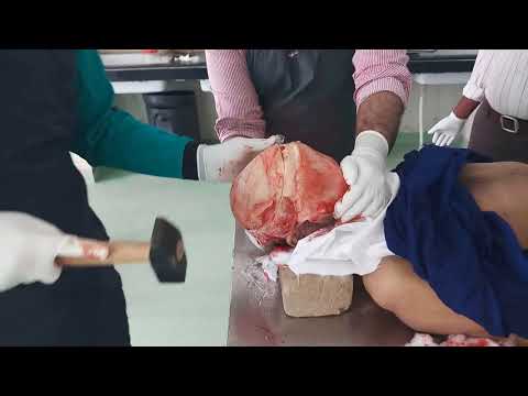
Opening the Skull Cap
- An outline for opening the skull cap (or vault) is made using an iron blade after retracting both flaps adequately. This involves marking anteriorly above the crown and around occipital areas.
- After outlining, a careful removal of the skull cap follows, exposing underlying structures such as dura mater and brain tissue beneath it. Dura mater removal aligns with previous incisions made during skull cap extraction.
Brain Removal Process
- The procedure includes cutting optic nerves on both sides while carefully handling surrounding structures like tentorium cerebelli, which must also be severed for complete brain removal.
- Once detached, both cerebellum sections are removed along with other brain components (pons and medulla), indicating successful extraction of soft brain tissue for fixation later on in formalin solution.
Abdominal and Chest Cavity Opening
Incision Techniques in Abdominal Surgery
- A new incision starts at the pubic symphysis extending towards sternal notch while sparing umbilicus; this deepens through various abdominal layers including superficial fatty layer and rectus sheath containing muscles underneath it.
- As layers are dissected downwards, identification of rectus muscles occurs leading towards peritoneum exposure where distinct smells indicate entry into abdominal cavity post-incision completion.
Chest Cavity Exploration
- Following abdominal work, attention shifts to chest cavity where similar dissection techniques apply; lengthening incisions allows easier access to underlying structures like ribs and sternocleidomastoid joint landmarks identified during exploration phase.
- Coastal cartilage cuts occur subsequently alongside sternal plate removals revealing pleural cavities housing lungs beneath them that show signs of adhesions due to prior inflammation or fluid accumulation within pleural space observed visually during surgery procedures conducted hereafter.
- These observations highlight potential complications arising from conditions affecting lung health such as pleural effusion noted throughout this segment's examination phase within thoracic region surgeries performed today.
Exploring the Pericardium and Heart Pathologies
Examination of the Pericardium
- The pericardium is examined, revealing an abnormal amount of fluid surrounding the heart, indicating potential pathologies.
- Notably, there is also increased fluid observed in the brain, suggesting systemic issues that may need further investigation.
Observations on Cardiac Condition
- Clotted blood is noted within the inferior vena cava during examination, which could indicate previous hemorrhagic events or complications.
- The heart appears enlarged with a fibrotic patch present; this suggests chronic conditions affecting cardiac function.
Lung and Liver Assessment
- Adhesions are observed in the lungs, indicating possible past infections or inflammatory processes that have led to structural changes.
- The liver is prioritized for removal during surgery to facilitate access to other organs.
Gastrointestinal Insights
- Upon inspection of the stomach, it contains liquefied contents rather than being empty; internal mucosa is visible.
- The small intestine's length and structure are demonstrated; its entry point can be cut for further exploration.
Large Intestine Features
- The large intestine shows distinct features such as haustra (pouches), which differentiate it from the small intestine.