Segunda Semana do Desenvolvimento (Embriologia Humana) - Bio Aulas
Introduction to Week Two of Embryology
Overview of the Second Week
- The video introduces the second week of embryological development, emphasizing that it will build upon concepts from the first week. A link to the previous video is provided for context.
- The focus is on visual learning rather than text-heavy materials; a summary and important questions are available in supplementary study material.
Key Events in Week Two
- Major events include differentiation of the trophoblast and embryoblast, leading to the formation of a bilaminar embryonic disc and completion of implantation.
- The blastocyst stage is revisited, highlighting its superficial connection with the endometrium at the end of week one.
Differentiation Processes
Trophoblast Differentiation
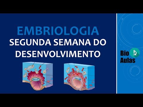
- In week two, the blastocyst fully implants into the endometrium. The trophoblast differentiates into two layers: syncytiotrophoblast and cytotrophoblast.
- Syncytiotrophoblast cells proliferate and invade the endometrium, forming a mass without distinct cell membranes, which contains multiple nuclei.
Cytotrophoblast Characteristics
- Remaining cells form cytotrophoblast; this differentiation results in two distinct layers within trophoblast structure.
- Syncytiotrophoblast acts like a cord pulling the blastocyst deeper into uterine tissue as it continues to grow.
Hormonal Production and Its Importance
Role of HCG
- Syncytiotrophoblast cells produce human chorionic gonadotropin (HCG), crucial for pregnancy detection through tests.
- HCG levels rise as gestation progresses due to increased production by growing syncytiotrophoblast cells.
Embryoblast Development
Formation of Bilaminar Disc
- The embryoblast also differentiates into two layers: epiblast (upper layer) and hypoblast (lower layer).
- Epiblast forms an amniotic cavity that will be lined with amniotic membrane producing amniotic fluid essential for embryo protection.
Development of the Embryo: Key Stages and Structures
Formation of Amniotic Fluid and Initial Differentiation
- The amniotic fluid is produced to protect the embryo, with initial differentiation of the trophoblast already underway. The syncytiotrophoblast is visible in pink, indicating early development.
Implantation Process
- As days progress, the syncytiotrophoblast invades the endometrium, leading to blastocyst implantation. Human Chorionic Gonadotropin (HCG) production increases during this phase. The cytotrophoblast surrounds the blastocyst as differentiation continues.
Bilaminar Embryonic Disc Formation
- By the second week, two layers form: epiblast (blue) and hypoblast (yellow), creating a bilaminar embryonic disc composed of these two cell layers. This structure is crucial for further development.
Cavities Forming Around the Embryo
- The amniotic cavity begins to develop, lined by amnioblasts that will produce amniotic fluid. Initially referred to as the blastocyst cavity, it transitions into what is now called the exocoelomic cavity or primitive yolk sac due to changes in nomenclature as structures evolve.
Nutritional Support through Maternal Blood
- As implantation progresses, lacunae form within the syncytiotrophoblast where maternal blood accumulates after vessels are breached. This process allows oxygen and nutrients from maternal blood to nourish the embryo via diffusion since it lacks its own vascular system at this stage. This mechanism is termed uteroplacental circulation.
Extraembryonic Mesoderm Development
- A region between the exocoelomic membrane and cytotrophoblast forms extraembryonic mesoderm with two types: splanchnic (adjacent to exocoelomic membrane) and somatic (next to cytotrophoblast). These areas begin developing isolated cavities that eventually merge into a single large chorionic cavity essential for further embryonic support structures like placenta formation.
Final Structure Overview
- The merging of cavities results in a significant space known as chorionic cavity; surrounding tissues include somatic extraembryonic mesoderm which contributes to future placental development—this area becomes critical for sustaining pregnancy and fetal growth throughout gestation periods.
Development of the Embryo: Key Structures and Stages
Formation of the Secondary Yolk Sac
- The future umbilical cord originates from a specific cavity, previously referred to as the primitive yolk sac. This cavity is now known as the secondary or definitive yolk sac after it forms and reduces in size.
- The exocoelomic membrane is a remnant that emerges from the primitive yolk sac, contributing to the formation of the secondary yolk sac.
Circulation Development
- By this stage, all lacunae within the structure are filled with blood, indicating the establishment of uteroplacental circulation.
Bilaminar Disc Structure
- At the end of week two, a bilaminar disc is formed consisting of two layers:
- Epiblast: Composed of tall columnar cells.
- Hypoblast: Made up of shorter cuboidal cells.
Precordal Plate Formation
- Towards the end of week two, certain hypoblast cells begin to resemble epiblast cells in a specific region, leading to the formation of what is called the precordal plate or oropharyngeal membrane.
- The precordal plate indicates where the mouth will develop in the embryo and marks an important cephalic region. Another structure called "membrane com clau" will also emerge at this time to indicate another significant area.