ANATOMÍA DE PELVIS Y PERINE
Anatomía de la Pelvis y del Periné
In this lecture, Dr. Miguel Ángel Estrada discusses the anatomy of the pelvis and perineum, highlighting their structures and functions.
Anatomy of the Pelvis
- The pelvis supports the upper body's weight and transfers it to the lower limbs, serving as an insertion point for various muscles.
- The pelvis protects and houses organs such as the bladder, rectum, uterus, and appendages while contributing to support, protection, and locomotion.
- The pelvis is divided into a false (greater) pelvis located superiorly and a true (lesser) pelvis situated inferiorly within the abdominal cavity.
Bones of the Pelvic Region
- The pelvic bones include ilium, pubis, and ischium which articulate with sacrum forming joints like sacroiliac joints.
- The ilium has distinct features like iliac crest for muscle attachment and spines for ligament connections.
- Pubis consists of body with superior & inferior rami forming structures like symphysis pubis & obturator foramen.
Detailed Bone Structures
- Ilium's anterior concave surface connects to muscles while posterior side has gluteal lines for muscle attachment.
- Pubis has a flattened body articulating with its counterpart via secondary cartilage at symphysis pubis forming a joint.
Understanding Pelvic Anatomy
In this section, the speaker delves into the intricate details of pelvic anatomy, discussing structures like the sacrum, pubis, and pelvis openings.
The Structure of the Pelvis
- The major sciatic spine and lesser sciatic spine are key components of the pelvis.
- The weight-bearing role of the psychiatric bone when sitting is highlighted.
Pelvic Openings and Formations
- Details about the upper pelvic ring being a closed ring formed by various structures like the promontory of the sacrum and sacral wings are discussed.
- The concept of pelvic narrowing during childbirth is explained in relation to passages for babies.
Different Types of Pelvis
- Four main types of pelvis are outlined: android, gynecoid, anthropoid, and platypelloid.
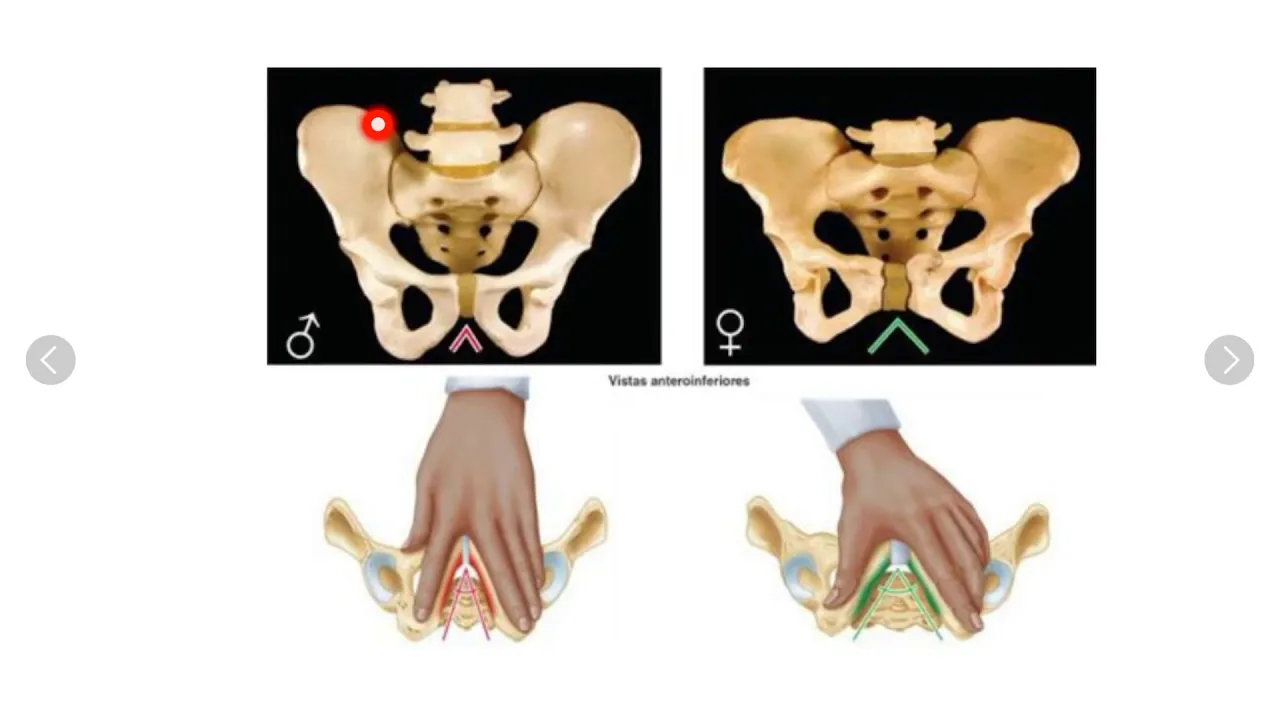
- Contrasts between male and female pelvises in terms of shape, weight-bearing capacity, angles, and dimensions are elucidated.
Importance of Pelvic Measurements
- The significance of measuring three narrowings in childbirth is emphasized: superior strait (pelvic inlet), mid-strait (interspinous diameter), and inferior strait (inter-tuberous diameter).
- Risks associated with narrow pelvic dimensions hindering childbirth are explored.
Obstetric Pelvis Anatomy
In this section, the anatomy of the obstetric pelvis is discussed, focusing on the various joints and ligaments that play a crucial role in childbirth.
Obstetric Pelvis Articulations
- The obstetric pelvis has multiple articulations between the last two lumbar vertebrae and the iliac bone through ligaments.
- The union between the iliac bone and sacrum forms the sacroiliac joint, consisting of anterior and posterior sacroiliac ligaments.
Ligaments and Joints
- Ligaments such as sacrotuberous and sacrospinous connect the sacrum to structures like ischium, forming a syndesmosis type of joint.
- Structures like pubic ligaments, symphysis pubis, longitudinal ligament, and sacrococcygeal ligament contribute to pelvic stability.
Sacroiliac Joint Details
- The anterior sacroiliac joint is synovial planar while the posterior one functions as a syndesmosis transferring energy to lower extremities.
- The acetabulum houses the femoral head with supporting structures like transverse ligament within its cavity.
Muscles in Pelvic Floor
- Muscles like levator ani (comprising puborectalis, pubococcygeus, iliococcygeus) form the pelvic floor providing support for pelvic organs.
Anatomy of the Pelvic Floor
In this section, the speaker discusses the anatomy of the pelvic floor, highlighting key muscles and structures in the region.
Muscles and Structures of the Pelvic Floor
- The pelvic floor includes muscles extending from the sacrum to the ischial spine, such as the obturator internus muscle. These muscles play a role in functions like urethra control and support for organs like the vagina.
- The levator ani muscle group comprises muscles like puborectalis, iliococcygeus, and pubococcygeus. These muscles contribute to anal and urethral function in both men and women.
- The obturator internus muscle runs from inside the obturator foramen to attach near the greater trochanter of the femur. It aids in hip stability and contributes to pelvic floor support.
Peritoneum and Female Reproductive Anatomy
This part delves into peritoneal structures in males versus females, focusing on their roles in reproductive anatomy.
Peritoneal Structures and Female Reproductive Anatomy
- The peritoneum covers various pelvic structures differently between males and females. In females, it forms a posterior cul-de-sac between rectum and uterus before continuing over bladder's roof towards anterior abdominal wall.
Anatomy of the Pelvis
In this section, the speaker delves into the anatomy of the pelvis, detailing structures and spaces within this region.
Structures in the Pelvic Region
- The area below includes the deep perineum and superficial perineal structures. Below the perianal membrane lies the superficial perineal space.
- Tabiques separate various structures in the pelvic region, such as ligaments connecting the bladder and pubis in women known as tubovesical ligaments.
Spaces and Ligaments in Pelvic Anatomy
- The presence of tabiques forms spaces like vesical spaces for circulation to the bladder, rectovesical spaces between rectum and sacrum.
- In men, prostatic tubes connect prostate and bladder while tabiques aid circulation to these organs.
Ligaments and Circulation in Pelvic Area
- Ligamentos útero sacro support pelvic stability. Circulation is mainly through six arteries in women and four in men.
- Arteries like gonadal arteries differ between genders with variations in their paths within the pelvis.
Arterial Supply to Pelvic Organs
In this section, the speaker discusses the arterial supply to various pelvic organs in detail.
Arterial Supply Details
- The umbilical artery branches into superior vesical arteries for upper bladder irrigation and uterine arteries for lateral cervical matrix supply.
- The ovarian artery arises from the anterior aorta, forming an ovarian tubal circuit for ovary irrigation. It can branch into vaginal arteries for lower bladder and vagina supply.
- The rectal artery supplies the middle rectum, while the pudendal artery exits through the greater sciatic foramen to irrigate the perineum and genitalia.
- The pudendal artery further provides blood flow to the lower rectum, perineum, labia majora, dorsal clitoris (in females), scrotum (in males), and dorsal penis (in males).
Venous Drainage Patterns in Pelvic Region
This part focuses on venous drainage patterns within the pelvic region.
Venous Drainage Insights
- Venous drainage involves vessels like superior vesical veins, uterine veins, vaginal veins, and middle rectal veins that typically drain into internal iliac vein or internal iliac artery region.
- Additional venous pathways include sacral lateral veins leading to vena cava directly and lumbar lymphatics connecting to common iliac vein or vertebral plexus internally.
Lymphatic Drainage in Pelvic Area
This segment delves into lymphatic drainage mechanisms specific to the pelvic area.
Lymphatic Drainage Summary
Anatomy and Physiology of the Pelvic Region
In this section, the speaker discusses the lymphatic drainage, innervation, and anatomical structures of the pelvic region.
Lymphatic Drainage
- The lymphatic drainage from the pelvic region flows towards the inguinal nodes.
- This drainage includes superficial inguinal nodes collecting from the perineum and deep inguinal nodes connecting to external iliac nodes.
- Ovarian lymphatic drainage primarily follows the ovarian plexus towards lumbar regions and sacral nodes.
- Metastasis in ovarian cancer can involve various lymph nodes necessitating their removal during surgery.
Innervation
- Nerve innervation in the pelvic region is mainly through lumbosacral plexuses.
- The sciatic nerve (L4-S3) innervates lower limbs with branches for specific muscles like piriformis and obturator internus.
- Visceral organs receive sympathetic innervation via mesenteric plexuses originating from lumbar plexuses.
- Sympathetic fibers increase tone in detrusor muscle for urine retention and anal sphincters for fecal continence.
Parasympathetic Innervation
- Parasympathetic nerves arise from S2-S4 forming pelvic splanchnic nerves.
- These nerves join to form hypogastric plexuses that reach pelvic organs, promoting relaxation of sphincters for micturition and defecation.
Anatomical Features of Perineum
This part delves into the anatomy of the perineum, highlighting its structure and functional significance.
Perineal Anatomy
- The perineum is a diamond-shaped area between thighs and buttocks, divided into urogenital anterior triangle and anal posterior triangle.
- It contains fibrous cartilaginous tissue known as perineal body or central tendon providing structural support.
Triangles of Perineum
- Anteriorly lies urogenital triangle bordered by pubis anteriorly, coccyx posteriorly, and tuberosities laterally.
- Posteriorly is the anal triangle with similar boundaries but enclosing anal canal or scrotum-vaginal junction based on gender.
Understanding Pelvic Anatomy
In this section, the speaker discusses the anatomy of the pelvis, focusing on structures such as the bladder, vagina, rectum, and prostate in males. The formation of various partitions within these regions is highlighted.
Structures and Partitions in Pelvic Anatomy
- The pelvis contains partitions separating the bladder from the vagina and the vagina from the rectum.
- In males, similar partitions form in regions like the prostatic region and between the rectum and bladder.
- Diafragmas pélvicos are discussed, including their fascia covering structures like obturador interno muscle and vaginal walls.
- The perineal membrane divides superficial and deep portions of perineum with contributions from muscles like obturador interno.
- Deep perineum or ischioanal fossa forms with contributions from muscles in the perineal region.
Understanding Pelvic Anatomy
In this section, the speaker delves into the anatomy of the pelvic region, discussing muscles like the compressor muscle of the urethra and various structures such as the external anal sphincter, bulbocavernosus muscles, and perineal membrane.
Muscles and Structures in the Pelvic Region
- The compressor muscle of the urethra is crucial in compressing the urethra.
- Various muscles include the external anal sphincter, transverse muscles, bulbocavernosus muscles, and ischiocavernosus muscles.
- The perineal membrane plays a role in supporting these structures.
Continuation of Pelvic Anatomy Discussion
This part continues to explore pelvic anatomy by focusing on fascial layers like Scarpa's fascia and deep structures such as bulbospongiosus muscles.
Fascial Layers and Muscles
- Scarpa's fascia extends into the fascia lata.
- Mention of CCOO phase formation involving muscular layers.
- Bulbospongiosus muscles originate from the perineal center towards ischiocavernosus bodies.
Nerves and Arteries in Pelvic Region
The discussion shifts towards nerves and arteries within the pelvic region, emphasizing their roles in innervation and blood supply.
Nerves and Arteries
- Nerves like pudendal nerves travel alongside internal pudendal arteries.
- Arteries branch off to supply regions like the urethra and dorsal penile artery.
Detailed Analysis of Anal Canal
Detailed examination of anatomical features within the anal canal including epithelial changes, vascular structures like hemorrhoidal vessels, lymphatic drainage patterns, and nerve innervation.
Anal Canal Features
- Transition from columnar to stratified epithelium at dentate line.
- Presence of hemorroidal vessels internally with lateral spaces for internal pudendal artery passage.
Vascular Supply in Pelvic Region
Focus on arterial pathways supplying rectum with details on branches originating from internal iliac artery towards rectum.
Vascular Pathways
- Internal pudendal artery exits pelvis via greater sciatic foramen before branching into rectal inferior artery.
New Section
The discussion revolves around the pudendal nerve originating from S2, S3, and S4. It touches upon the practice of episiotomy during childbirth and its declining use due to associated risks.
Pudendal Nerve and Episiotomy
- The pudendal nerve arises from S2, S3, and S4.
- Episiotomy involves a cut in the vaginal introitus region during childbirth but is now less common due to risks like perineal body cuts leading to anal region tears and fistula formation.