CAP 51 4/4: Células horizontales, bipolares, amacrinas y ganglionares l Fisiología de Guyton
Welcome and Overview
The video discusses the receptor function in the retina, detailing the cells involved in this process.
Retina Structure and Cell Functions
- The retina consists of various layers including the pigment layer, photoreceptor layer housing rods and cones, external nuclear layer with rod and cone nuclei, external plexiform layer for synapses, and more.
- The internal nuclear layer contains bipolar cell nuclei while the internal plexiform layer involves synapses between bipolar cells and amacrine or ganglion cells.
- Rods and cones hyperpolarize to initiate electrical conduction. Horizontal cells facilitate lateral inhibition by conducting electricity horizontally between cones.
Bipolar Cells and Signal Transmission
- Bipolar cells conduct signals from horizontal cells towards ganglion cells in an anterograde manner. They can form synapses with amacrine or ganglion cells.
- Amacrine cells can conduct electricity horizontally or vertically towards ganglion cells to transmit impulses generated by rods or cones.
Ganglion Cells and Impulse Transmission
- Ganglion cells transmit impulses to the optic nerve. Interplexiform neurons inhibit synapses in the external plexiform layer retrogradely.
- Not all impulses follow the same path; transmission speed varies based on peripheral versus central retinal vision types (rods vs. cones).
Retinal Vision Types
Contrasts peripheral retinal vision dominated by rods with central retinal vision rich in cones for faster impulse transmission.
Central Retina Characteristics
- Central retina has abundant cones leading to rapid signal transmission through bipolar to ganglion cells.
- Horizontal and amacrine cell functions include lateral inhibition. Peripheral retina has both rods and cones but emphasizes rod function for signal transmission efficiency.
Signal Pathways Comparison
- In central retina, each cone connects directly to a bipolar cell then a ganglion cell for efficient impulse transmission compared to peripheral retina's indirect pathway via single bipolar cell per multiple rods.
- Cones have dedicated bipolar and ganglion cells per cone, unlike rods sharing resources among multiple rods in peripheral vision.
Neurotransmitters in Retina
Explores neurotransmitter release by retinal neurons beyond nervous impulses for synaptic communication enhancement.
Neurotransmitter Release Mechanisms
Detailed Analysis of Retinal Cells and Neurotransmitters
In this section, the discussion revolves around the neurotransmitters released by retinal cells, focusing on the uncertainty regarding the specific substances released by certain cells and highlighting the well-understood neurotransmitters like glutamate released by rods and cones.
Neurotransmitter Release by Different Retinal Cells
- Some retinal cells release neurotransmitters whose identity remains uncertain.
- Rods and cones are known to release glutamate when inactive.
- Bipolar, horizontal, and amacrine cells release inhibitory neurotransmitters such as GABA, glycine, dopamine, acetylcholine, and indolamine.
Electrical Conduction in Retinal Cells
- Electrotonic conduction is emphasized over action potentials for signal transmission in retinal cells.
- Electrotonic conduction occurs mainly in dendrites through membrane potentials that do not trigger action potentials but stimulate ganglion cells eventually.
Action Potentials vs. Electrotonic Conduction
- Electrotonic conduction leads to graded responses regardless of stimulus intensity compared to all-or-nothing action potentials.
- Action potentials require a strong stimulus to generate a response compared to electrotonic conduction's continuous flow of electricity.
Role of Ganglion Cells in Signal Transmission
- Ganglion cells are crucial for transmitting signals via action potentials within the retina.
- Only ganglion cells consistently use action potentials for signal transmission within retinal neurons.
Functionality of Horizontal Cells in Image Processing
- Horizontal cells primarily facilitate lateral inhibition in response to light stimuli, contributing to image contrast enhancement.
- Inhibition generated by horizontal cells prevents excessive signal propagation between photoreceptors, aiding visual contrast perception.
Insights into Bipolar Cells' Response Mechanisms
This segment delves into the dual nature of bipolar cells as either excitatory (depolarizing/on-cells) or inhibitory (hyperpolarizing/off-cells), elucidating their distinct responses based on glutamate levels and light conditions.
Classification of Bipolar Cells
- Bipolar cells can be categorized as either depolarizing/excitatory or hyperpolarizing/inhibitory based on their response mechanisms.
Glutamate Influence on Bipolar Cell Activity
- Glutamate levels play a pivotal role in modulating bipolar cell activity under varying light conditions.
Excitatory vs. Inhibitory Responses
Understanding Retinal Cells and Functions
In this section, the discussion revolves around the role of different types of retinal cells, focusing on how they respond to light stimuli and contribute to visual processing.
Glutamate's Role in Cell Excitation and Inhibition
- Glutamate affects cell excitability differently based on cell type.
- Sodium and calcium channels are blocked by glutamate in excitatory cells.
- Glutamate opens sodium and calcium channels in inhibitory cells, activating them.
Factors Influencing Bipolar Cell Activity
- Bipolar cells' activity is influenced by direct and indirect stimulation.
- Glutamate can stimulate horizontal cells, indirectly inhibiting excitatory cells.
- Inhibitory bipolar cells are directly stimulated by glutamate.
Light Response Mechanisms in Bipolar Cells
- Light exposure triggers changes in bipolar cell activity.
- Decreased glutamate release during light exposure activates previously inhibited excitatory cells.
- This shift leads to positive signals from excitatory cells and negative signals from inhibitory ones.
Exploring Amacrine Cells in Retina
The focus shifts to amacrine cells within the retina, detailing their functions and classifications based on morphology and histochemistry.
Diversity of Amacrine Cells
- Over 30 classes of amacrine cells have been identified through morphological analysis.
- Functions of some amacrine cell types include involvement in direct vision pathways with rods, cones, bipolar, and ganglion cells.
Activation Patterns of Amacrine Cells
- Different amacrine cell types respond uniquely to visual stimuli patterns.
- Some amacrine cells activate during continuous visual signals while others respond when signals cease or change intensity.
Specialized Responses of Amacrine Cells
- Specific amacrine cell subtypes exhibit distinct response patterns to light variations.
- Some amacrines rapidly respond then inhibit upon stimulus cessation; others only activate with light onset or offset.
Role of Macular Ganglion Cells
Delving into ganglion cells located at the macula region, highlighting their significance as conduits for visual information transmission towards the brain.
Functionality of Ganglion Cells
- Macular ganglion cells play a crucial role in transmitting retinal information to the brain for further processing.
Detailed Analysis of Retinal Ganglion Cells
In this section, the speaker delves into the characteristics and functions of retinal ganglion cells, focusing on their distribution in the retina and their roles in visual processing.
Characteristics of Peripheral Retina
- Each ganglion cell in the periphery receives input from approximately 200 rods, making the peripheral retina highly sensitive to dim light.
- Stimulation of one rod among these 200 leads to an electrotonic current that activates ganglion cells, particularly in dark conditions.
Types of Ganglion Cells
- Initial studies on cat retinas identified cells W, X, and Y as significant ganglion cell types.
- Cells W are slow-speed cells connected to rods, primarily involved in low-light vision.
Functions of Different Ganglion Cell Types
- Cells X have small detection fields associated with cones for color vision and high-speed processing.
- Cells G possess a high speed of 50 meters per second and detect rapid visual changes, aiding eye movements towards stimuli.
Continuation: Retinal Ganglion Cell Classification
This section explores further classifications of retinal ganglion cells based on their characteristics and connections within the visual pathway.
Main Classes of Ganglion Cells
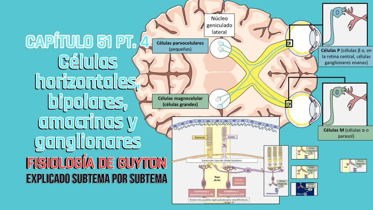
- The most crucial ganglion cell types found in primates and humans are P (beta) and M (alpha/parasol), each with distinct features.
- These cells project to specific regions in the brain; P cells terminate at parvocellular neurons while M cells end at magnocellular neurons.
Pathway to Visual Cortex
- Axons from P cells reach the lateral geniculate nucleus's parvocellular group, whereas M cell axons connect with magnocellular neurons.
- Parvocellular neurons are more lateral while magnocellular neurons are ventral within the lateral geniculate nucleus.
Insights into Ganglion Cell Receptors
This segment focuses on receptor characteristics of different ganglion cell types and their specialized functions in visual perception.
Receptor Characteristics
- P cells have small receptive fields for fine details and slow conduction but sustained responses to colors.
Understanding the Role of Ganglion Cells in Vision
In this section, the focus is on the role of ganglion cells in vision and how they respond to light stimuli.
The Significance of Ganglion Cells
- Different types of retinal cells like cones, rods, horizontal cells, bipolar cells, and ganglion cells have distinct functions.
- Ganglion cells exhibit continuous impulses even without stimulation, crucial for long-distance electrical conduction.
- Discussion on how ganglion cells respond to changes in light intensity through firing patterns.
Neural Mechanisms Behind Light Detection
This part delves into the neural mechanisms involved in detecting light and its impact on ganglion cell activity.
Light Detection and Ganglion Cell Response
- Ganglion cells show continuous impulses even in darkness but decrease glutamate release when exposed to light.
- Detailed explanation of how ganglion cell stimulation changes with varying light conditions.
- Activation of ganglion cells by light leads to increased nerve impulses transmission.
Inhibition Mechanisms in Retinal Processing
Exploring inhibitory mechanisms within retinal processing and their effects on ganglion cell activity.
Inhibition Effects on Ganglion Cells
- Horizontal and amacrine cells play a role in lateral inhibition affecting neighboring ganglion cell activity.
- Inhibitory lateral effects lead to contrast enhancement by modulating ganglion cell responses based on light intensity variations.
Color Signal Processing in Retina
Understanding how color signals are processed within the retina involving cone interactions with bipolar and ganglion cells.
Color Signal Processing
- Bipolar off-cells get activated by glutamate release under dark conditions, influencing ganglionic cell responses.
- Uniform illumination across the retina results in no specific contrast generation due to balanced inhibitory effects.
Detailed Explanation of Retinal Processing
In this section, the speaker delves into the intricate process of retinal processing, focusing on how bipolar cells are connected to ganglion cells and the role of different types of cones in color detection.
Bipolar Cells and Ganglion Cells Connection
- Bipolar cells are linked to only one ganglion cell.
- The absence of light stimuli results in the non-activation of cones.
Role of Glutamate and Cone Activation
- Glutamate inhibits the ON cell that should stimulate another cell.
- Removal of glutamate due to cone activation leads to bipolar cell activation, subsequently activating ganglion cells.
Color Detection Mechanisms in Retina
This part elucidates how specific wavelengths reaching individual cones trigger distinct responses in bipolar cells, influencing color perception and contrast detection.
Cone Specific Wavelength Stimulation
- When a single wavelength reaches a specific cone (e.g., red wavelength for red cone), it gets stimulated.
- Other bipolar cells generate direct inhibition on ganglion cells based on stimulation from different colored rods.
Significance of Color Contrast Mechanisms
The importance of color contrast mechanisms within retinal processing is discussed, emphasizing their role in initiating color distinction at the retinal level rather than in the brain.
Color Contrast Detection
- Red rod excitation and green/blue rod inhibition lead to ganglion cells perceiving contrast.