Mesencephalon (Midbrain) - External & Internal structures + QUIZ | Anatomy
Introduction to the Anatomy of the Central Nervous System
In this section, Meditay introduces the anatomy of the Central Nervous System, specifically focusing on the Midbrain or mesencephalon.
Anatomy of the Midbrain
- The central nervous system consists of two parts: the encephalon and the spinal cord.
- The encephalon is further divided into specific parts, including the brainstem (Medulla, Pons, and midbrain), cerebellum, Diencephalon, and telencephalon.
- The focus of this video is on the Midbrain.
- The Midbrain is located above Pons, in front of the Cerebellum, and below the Diencephalon.
External Surfaces of the Midbrain
This section covers the external surfaces of the midbrain from an anterior view and a posterior view.
Anterior Surface
- The majority of the anterior surface of the midbrain is occupied by Cerebral Peduncles.
- Cerebral Peduncles are large peduncles responsible for voluntary movement and contain tracts from the Cortex of your cerebrum.
- Within interpeduncular Fossa (located between cerebral peduncles), structures like Hypothalamus and Pituitary gland can be found. However, they are not part of the midbrain.
Posterior Surface
- From a posterior view, significant structures associated with midbrain include cerebral peduncles and Tectal Plate or Lamina Tecti.
- On Tectal Plate, there are four rounded structures known as superior colliculus responsible for rapid eye movements.
- Fibers from Lateral Geniculate bodies go to superior colliculi through Brachium of the superior colliculi, activating the tectospinal tract for coordinated eye and neck muscle movements.
- Inferior colliculi, located under the superior colliculi, are part of the hearing pathway.
Structures of the Midbrain
This section focuses on specific structures within the midbrain.
- The posterior perforated substance is a depression on the anterior surface of the midbrain containing grey matter and small holes for blood vessels.
- The ocular motor sulcus of the mesencephalon is a groove through which the oculomotor nerve exits. This nerve innervates extrinsic eye muscles responsible for eye movement.
- Other structures associated with the midbrain include interpeduncular Fossa, Hypothalamus, and Pituitary gland.
Visual Pathway and Motor Impulses
This section explains how visual information is processed in relation to the midbrain.
- Optic nerves carry signals from retinas to optic chiasm where fibers cross over.
- Lateral geniculate bodies receive fibers from optic chiasm and send them to occipital lobe's primary visual cortex for conscious awareness.
- Fibers also go from lateral geniculate bodies to superior colliculi via Brachium of superior colliculi. This activates tectospinal tract for coordinated eye and neck muscle movements.
- Cochlear nerve carries sound impulses from cochlea to cochlear nuclei in Pons, which is part of the hearing pathway.
Timestamps are approximate and may vary slightly depending on video playback.
New Section
This section provides an overview of the external surface of the midbrain.
External Surface of the Midbrain
- The medial geniculate body sends impulses to the primary auditory cortex in the primary temporal gyrus.
- The lateral sulcus of the mesencephalon is located on the posterior surface, serving as a border between the cerebral peduncles and the posterior surface of the midbrain.
- The trigone of the lateral lemniscus is found in this area.
- Below the inferior colliculus, cranial nerve number 4 (trochlear nerve) innervates the superior oblique muscle of the eye.
New Section
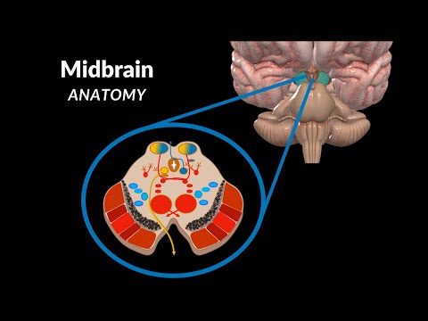
This section explores a cross-section view of the midbrain at the level of superior colliculi.
Cross-section View at Superior Colliculi
- Anteriorly, there are cerebral peduncles with interpeduncular fossa in between.
- The aqueduct of the midbrain connects the third ventricle with the fourth ventricle as part of the ventricular system.
- The internal surface can be divided into three regions: tectum (containing colliculi), tegmentum, and cerebral peduncles.
New Section
This section focuses on structures within grey matter and their functions.
Grey Matter Structures
- The red nucleus or nucleus ruber is pale pink in color due to iron content. It contributes to coordinating voluntary muscle control through extrapyramidal pathways.
- Substantia nigra consists of two distinct regions: pars compacta and pars reticulata. Pars compacta contains dopamine neurons producing neuromelanin and plays a significant role in basal ganglia function.
New Section
This section provides an overview of the internal structures of the midbrain.
Internal Structures
- The internal surface can be divided into three regions: tectum, tegmentum, and cerebral peduncles.
- Grey matter contains nuclei of neurons, while white matter consists of myelinated nerve fibers.
- The basal ganglia, including the substantia nigra, play a role in coordinating motor functions through communication with the primary motor cortex.
New Section
This section explains the role of basal ganglia in motor coordination.
Basal Ganglia and Motor Coordination
- Basal ganglia consist of grey matter nuclei within the brain, including caudate nucleus, putamen, globus pallidus, thalamus (ventral anterior and ventral lateral parts), subthalamic nuclei, and substantia nigra.
- Basal ganglia modify motor plans from the primary motor cortex before sending them back to initiate or modulate movement.
- The nigrostriatal pathway uses dopamine to start, stop, and modulate movement.
New Section
This section highlights the communication between primary motor cortex and basal ganglia for coordinated movement.
Communication Between Primary Motor Cortex and Basal Ganglia
- The primary motor cortex sends motor tracts along the spinal cord via the corticospinal tract to engage muscles for conscious limb movements.
- To execute proper motor plans, communication with basal ganglia is necessary. Basal ganglia modify and send back modified plans to initiate or modulate movement.
Timestamps are provided for each section based on available information in the transcript.
Starting Point in Studying Midbrain Physiology
This section provides an introduction to studying the physiology of the midbrain and discusses the nuclei found in different layers.
Grey Matter Nuclei
- The superior colliculi level contains the nucleus of the oculomotor nerve and the posterior accessory nucleus of the oculomotor nerve. The oculomotor nerve innervates extraocular muscles for eye movement, while the posterior accessory nucleus provides parasympathetic innervation to the eyes.
- At the inferior colliculi level, there is the nucleus of the trochlear nerve, which supplies the superior oblique muscle for eye movement.
- The mesencephalic nucleus of the trigeminal nerve is found at the level of Inferior colliculus and receives sensory information from muscles of mastication. Other nuclei of trigeminal nerve include principal nucleus (touch and vibration), spinal nucleus (pain and temperature), and motor nucleus (motor innervation for muscles of mastication).
- The reticular formation is essential for vital functions and balance, while periaqueductal grey substance is associated with pain reduction.
White Matter Tracts in Midbrain
This section focuses on white matter tracts in the midbrain, including ascending sensory tracts and descending motor tracts.
Ascending Tracts
- The medial lemniscus is an ascending tract that carries sensory fibers related to conscious proprioception and mechanoreceptors from lower parts (gracile fascicle) and upper parts (cuneate fascicle) of the body to reach primary somatosensory area in cerebral cortex.
- The spinal lemniscus is formed by the anterior and lateral spinothalamic tracts, which join together between the pons and medulla. It carries conscious sensory input related to pain, temperature, pressure, and touch to the primary somatosensory area in the cortex.
- The trigemnical lemniscus receives sensory input from the facial area through the trigeminal ganglion. Fibers cross to the other side in the pons and ascend through the midbrain to reach the primary somatosensory area.
- The lateral lemniscus is part of the hearing pathway, carrying impulses from cochlear nuclei in Pons to inferior colliculi via trapezoid body. From there, impulses are sent to medial geniculate body and then to primary auditory cortex in superior temporal gyrus.
Descending Tracts in Midbrain
This section discusses descending tracts found along cerebral peduncles and tegmentum of the midbrain.
Tegmentum
- The posterior tegmental decussation is a tract that originates from superior colliculus and decussates within tegmentum.
The transcript does not provide further information on descending tracts along cerebral peduncles.
Please note that this summary only covers a small portion of the video transcript provided.
Descent of Tracts in the Midbrain
This section discusses the descent of tracts in the midbrain, specifically focusing on the optic chiasm, lateral geniculate bodies, superior colliculi, and tegmental decussations.
Optic Chiasm and Lateral Geniculate Bodies
- The optic nerves cross at the optic chiasm and synapse with the lateral geniculate bodies.
- Fibers from the lateral geniculate bodies go back to the occipital lobe for conscious perception of visual stimuli.
- Fibers also go from the lateral geniculate bodies to the superior colliculi.
Superior Colliculi and Tectospinal Tract
- Fibers from the superior colliculi form the tectospinal tract.
- Engagement of the superior colliculi activates neck muscles based on visual stimuli.
Anterior Tegmental Decussation
- The rubrospinal tract forms an anterior tegmental decussation as it leaves the red nuclei.
Corticopontine Tract and Pontocerebellar Tract
- The corticopontine tract synapses with pontine nuclei in Pons after originating from the cortex.
- Fibers then travel to the cerebellum as pontocerebellar tracts.
- From there, they reach nucleus ruber as cerebellorubral tracts.
Reticulospinal Tract and Medial Longitudinal Fasciculus
- The reticular formation in the brainstem gives rise to reticulospinal tracts responsible for balance and posture control.
- The medial longitudinal fasciculus coordinates involuntary movements of head, neck, and eyes through cranial nerve synapses.
Descending Tracts in Cerebral Peduncles
This section focuses on the descending tracts within the cerebral peduncles, including the corticospinal tract, corticonuclear tracts, and corticopontine tract.
Corticospinal Tract
- The corticospinal tract originates from pyramidal cells in the primary motor area.
- It descends to the spinal cord to innervate skeletal muscles.
Corticonuclear Tracts
- Corticonuclear tracts are responsible for voluntary control of muscles in the head and neck.
- They descend alongside the corticospinal tract.
Corticopontine Tract
- The corticopontine tract synapses with pontine nuclei in Pons within the cerebral peduncle.
- Fibers then travel to the cerebellum as pontocerebellar tracts.
Summary of Mesencephalon
This section provides a summary of all the grey and white matter structures discussed in the mesencephalon.
- A table is presented with names of nuclei and tracts along with descriptions.
- The viewer is encouraged to identify each structure based on the provided information.
Timestamps have been associated with relevant bullet points.