PLEXO SACRAL E COCCÍGEO
Plexo Sacral e Coccígeo: Estruturas e Funções
Introdução ao Plexo Sacral
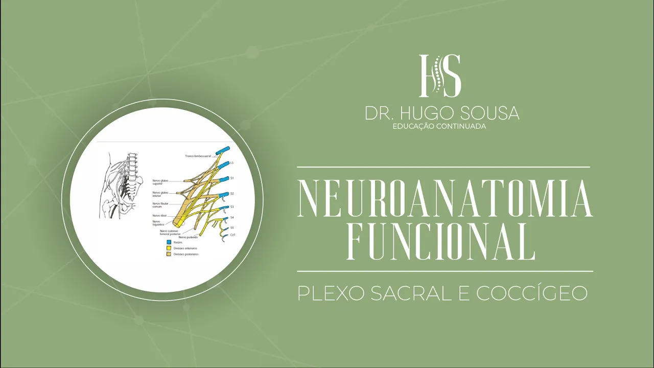
- O plexo sacral está localizado imediatamente inferior ao plexo lombar, conforme ilustrado na imagem apresentada.
- É formado por ramos anteriores dos nervos espinhais L4 a S3, com alguns autores incluindo o L5.
- Os nervos originados do plexo sacral inervam a parte inferior do dorso, a face posterior da coxa, a perna e as regiões dorsal e plantar do pé.
Estrutura do Plexo Sacral
- O plexo sacral é dividido em divisões anteriores e posteriores, que contêm fibras dos nervos lombares.
- As raízes que formam o plexo são L4, L5, S1, S2 e S3; destacando-se o nervo isquiático como um dos mais espessos do corpo humano.
Nervo Isquiático
- O nervo isquiático é misto e possui raízes de L4 a S3; ele inerva partes da perna até o pé.
- A união das raízes L4 e L5 forma um tronco chamado tronco lombossacral.
Ramos Importantes do Plexo Sacral
- Os principais ramos incluem os nervos glúteos superior e inferior, além do nervo isquiático.
- Esses nervos têm funções significativas relacionadas à inervação muscular na região glútea.
Ramos Musculares
- Os ramos musculares incluem aqueles para os músculos piriforme, quadrado femoral e gêmeos inferiores.
- Outros importantes são os nervos para o obturador interno e gêmeos superiores; todos estão relacionados à divisão anterior.
Nervos Cutâneos e Funcionalidade
- O nervo cutâneo femoral posterior é um componente misto importante no plexo sacral.
- O nervo pudendo é crucial para a inervação da pele na região pélvica; também se destaca pela sua função na continência fecal e urinária.
Conclusão sobre o Plexo Coccígeo
- O ramo perineal do quarto sacral contribui para a funcionalidade anal; enquanto o nervo ano coccígeo pertence ao plexo coccígeo.
Nerve Anatomy and Innervation of the Lower Limb
Overview of Nerve Distribution
- The discussion begins with the identification of various nerves in the lower limb, including those for the femoral quadriceps and superior/inferior gemelli.
- It highlights mixed contributions from anterior and posterior divisions, mentioning common fibular nerve roots L4-S2 and S3.
Pathways of Nerves
- Nerves traverse foramina to exit the pelvic cavity, either through the greater sciatic foramen or lesser sciatic foramen.
- The obturator nerve is noted as it passes beneath the inguinal ligament into the thigh.
Specific Nerve Functions
- The superior gluteal nerve (L4-L5-S1) is responsible for innervating muscles involved in hip abduction, such as gluteus minimus and medius.
- The inferior gluteal nerve (L5-S2) innervates the gluteus maximus, crucial for hip extension.
Muscle Actions and Innervation
- Discussion on muscle actions includes external rotation by piriformis and abduction by gluteal muscles.
- The quadratus femoris and gemelli are also identified as external rotators with specific nerve innervations from L4-L5-S1.
Cutaneous Innervation
- Cutaneous branches provide sensory innervation to specific skin areas; notably, S2-S3 contributes to sensation in medial buttock regions.
- The cutaneous femoral posterior nerve's territory includes areas like the posterior thigh and part of the perineum.
Sciatic Nerve Significance
- The sciatic nerve (L4-S3), composed of tibial and common fibular nerves, plays a vital role in lower limb function.
Nerve Anatomy and Innervation
Overview of Popliteal Nerve Branches
- The popliteal nerve divides into two branches: the common fibular nerve (lateral) and the tibial nerve (medial), responsible for innervating specific myotomes.
Myotome Innervation
- The first myotome discussed is the semitendinosus and semimembranosus muscles, along with the adductor magnus in the thigh, which receives innervation from both the sciatic nerve and obturator nerve.
Tibial Nerve Functions
- The tibial nerve innervates several structures including:
- Skin on the posterior surface of the leg and sole of the foot.
- Muscles such as gastrocnemius (both medial and lateral heads), plantaris, popliteus, soleus, flexor digitorum longus, tibialis posterior, and flexor hallucis longus.
Sensory Innervation by Tibial Nerve
- The tibial nerve also provides sensory innervation through its cutaneous branches:
- Originates sural nerve affecting sensitivity on posterior surface and part of anterior-lateral foot.
- Includes calcaneal branches contributing to this sensory territory.
Common Fibular Nerve Overview
- The common fibular nerve primarily serves:
- Sensitivity to dorsal foot area and anterolateral surface of the leg.
- Motor functions include innervating muscles like fibularis longus, fibularis brevis, tibialis anterior, extensor hallucis longus, extensor digitorum longus, extensor hallucis brevis, and extensor digitorum brevis.
Deep vs. Superficial Fibular Nerves
- The common fibular nerve splits into:
- Deep fibular nerve: Innervates tibialis anterior and extensors.
- Superficial fibular nerve: Responsible for innervating lateral compartment muscles (fibularis longus & brevis).
Clinical Relevance of Fibular Nerve
- Importance in surgical procedures; superficial nature makes it susceptible to injury near head of fibula.
- Palpation can reveal sensations indicating potential issues with this peripheral structure.
Consequences of Injury to Fibular Nerve
- Damage leads to characteristic gait changes due to paralysis in anterior compartment muscles resulting in a "foot drop" condition where individuals drag their toes while walking.
Surgical Applications
Anatomy and Function of the Pudendal Nerve
Dissection and Observations
- The dissection reveals the anterior and lateral aspects of the leg, highlighting a simple extension that allows for its resection and use in grafting.
- Post-surgery, patients may experience paresthesia or anesthesia in specific territories due to nerve involvement.
Pudendal Nerve Anatomy
- The pudendal nerve branches into areas including the perianal region and urogenital area, reinforcing its role in innervating other nerves like the cutaneous femoral nerve.
- It innervates various muscles such as the deep transverse perineal muscle, external urethral sphincter, and provides sensory input to genital regions.
Functional Implications
- The pudendal nerve is both sensory and motor; it plays a crucial role in pelvic floor function.
- Its relationship with other structures like the sacrotuberous ligament aids in understanding pudendal nerve block locations.
Clinical Applications
- Understanding anatomical landmarks near the ischial tuberosity can guide effective pudendal nerve blocks for pain management.
- Anesthetic injections can block pudendal nerve action, impacting sensation and motor control in targeted areas.
Pelvic Splanchnic Nerves
- Pelvic splanchnic nerves (S2-S4 roots) have both sensory functions related to pelvic organs and motor functions affecting visceral activities.
- These nerves are part of the parasympathetic system, differentiating from sympathetic pathways which are located near vertebral bodies.
Role in Sexual Function
- Pelvic splanchnic nerves stimulate erection by increasing blood flow to erectile tissues; they also modulate digestive tract mobility.
- They play a significant role in sexual arousal mechanisms for both males (penis) and females (clitoris), emphasizing their importance in reproductive health.
Impact on Urinary Function
- These nerves influence bladder detrusor muscle activity, facilitating urination while inhibiting internal urethral sphincter contraction.
- Damage to pelvic splanchnic nerves can lead to urinary retention issues post-surgery, exemplified by cases following prostatectomy procedures.
Surgical Considerations
Understanding the Pelvic Floor and Its Nerves
Anatomy of the Pelvic Region
- Discussion on how prostate lesions can lead to erectile dysfunction, highlighting the role of visceral sensory afferents that follow parasympathetic pathways from pelvic viscera.
- Introduction of the nerve responsible for innervating the levator ani muscle, which is crucial for pelvic floor function, including its connection to other muscles like the coccygeus.
- Explanation of the importance of pelvic floor muscles in supporting pelvic organs and their anatomical relationships with surrounding structures.
Sensory Functions and Reflexes
- Description of sensory functions related to a small area between the anus and coccyx, emphasizing its contribution to overall pelvic health.
- Overview of the coccigeal plexus, detailing its contributions from sacral roots (S4-S5), which are essential for innervating perianal skin and reflex actions.
Clinical Relevance
- The coccigeal nerve's role in anal reflexes is discussed, particularly how mechanical stimulation can trigger muscle contractions in response to sensory input.
- Mention of additional sacral roots involved in pelvic floor interactions, reinforcing their significance in clinical assessments and interventions.
Summary of Key Structures
- Clarification on how coccigeal nerves penetrate pelvic floor muscles and ligaments to innervate perineal skin areas effectively.