ACTIVIDAD ELECTRICA SIN PULSO
Activity of Electrical Impulse in Clinical Cases
Introduction to the Case Study
- Walter Sazo introduces the topic of electrical impulse activity in clinical settings, emphasizing its relevance for future medical professionals.
- The session begins with a clinical case to provide context for understanding emergency situations encountered in hospitals.
Clinical Case Overview: José
- José, a 28-year-old male security guard, presents with no significant medical history but has sustained two gunshot wounds to the abdomen during a robbery.
- Upon arrival at the hospital, he is conscious but exhibits signs of shock: pale skin, sweating (diaphoresis), and intense abdominal pain with active bleeding. Initial vital signs show low blood pressure (90/60) and high heart rate (130 bpm).
Initial Evaluation Protocol
- Emphasis on performing an initial evaluation using the ABCDE approach: Airway, Breathing, Circulation, Disability, Exposure. This systematic assessment is crucial in emergencies.
- The patient’s airway is clear; however, breathing is inadequate with only eight breaths per minute and oxygen saturation at 85% despite supplemental oxygen. Circulation shows cold and pale skin without detectable radial pulse but weak carotid pulse. Neurological status indicates unconsciousness. Two penetrating wounds are noted on examination.
Importance of Continuous Assessment
- Reinforcement of the need for continuous monitoring during patient evaluation and treatment; students should observe practices by residents or interns during their training periods in hospitals. Always prioritize ABCDE assessments when managing patients in critical conditions.
Cardiac Activity Analysis
- The patient does not exhibit spontaneous breathing or palpable pulses; an ECG may show sinus rhythm despite clinical signs indicating cardiac arrest due to lack of detectable pulse—this condition is termed "pulseless electrical activity."
- Understanding that while there may be organized electrical activity visible on monitors, it does not equate to effective cardiac contractions or perfusion—critical knowledge for emergency responders.
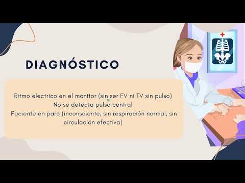
Defining Pulseless Electrical Activity
- Pulseless electrical activity (PEA) refers to a situation where there are organized heart rhythms present on an ECG monitor without corresponding mechanical contractions leading to blood flow—an important distinction for diagnosis and treatment strategies in emergencies.
- Key physiological factors necessary for effective cardiac function include proper electrical activity generation from pacemaker cells, structural integrity of myocardial tissue, adequate preload and afterload conditions as well as sufficient coronary perfusion pressure—all essential components that can affect outcomes in cases like José's.
Understanding Cardiac Function and Mechanisms
Key Concepts of Cardiac Contraction
- The effectiveness of cardiac contraction relies on the presence of electrical impulses; any disruption can prevent proper depolarization and mechanical contraction, leading to inadequate blood flow.
- Important physiological mechanisms include the "Hs" and "Ts," which are critical for understanding cardiac function. These should be recalled when associating various concepts related to heart mechanics.
Electromechanical Dissociation
- Electromechanical dissociation occurs when myocardial depolarization happens without effective contraction, often due to energy depletion (e.g., ATP deficiency) or severe myocardial damage such as ischemia or hypoxia.
- Conditions like severe acidosis or hypoxia can lead to myocardial ischemia, impacting the heart's ability to contract effectively. Examples include injuries from infarction or other physiological causes that compromise blood supply.
Impact of Preload on Cardiac Output
- Alterations in preload (ventricular filling) can occur even if the heart is electrically active; insufficient volume prevents effective blood ejection, particularly in cases of severe hypovolemia due to active hemorrhage.
- According to Frank-Starling's law, increased venous return enhances cardiac contractility; however, significant blood loss results in inadequate preload despite ongoing cardiac activity, leading to a lack of palpable pulse.
Mechanical Obstruction and Contractility Issues
- Mechanical obstructions (e.g., pulmonary embolism) can block blood flow while maintaining electrical activity within the heart; this highlights the distinction between electrical signals and actual perfusion capability.
- Effective contractility may be compromised by conditions such as hypoxia, acidosis, hyperkalemia, or overdose from myocardial depressants like beta-blockers or calcium antagonists. Understanding patient history is crucial for diagnosis and treatment planning.
Clinical Diagnosis and Treatment Approaches
- In cases where patients exhibit signs of cardiac arrest without detectable pulses, clinicians must assess both clinical signs and monitor rhythms using defibrillation equipment to identify non-shockable rhythms like pulseless ventricular tachycardia or asystole.
- Initiating CPR is essential for patients in cardiac arrest with non-defibrillable rhythms; airway management should follow basic life support protocols while ensuring intravenous access for fluid resuscitation in cases of hypovolemia. Advanced airway devices may be necessary if standard methods fail.
ACLS Protocols and Administration of Medications
Intravenous Access and Medication Administration
- Intravenous access is crucial for administering crystalloids and medications during ACLS, facilitating effective patient management.
- Vasopressin is less commonly used in hospitals; however, epinephrine (adrenaline) is the primary medication administered during cardiac arrest situations.
- Epinephrine should be given at a dose of 1 mg every 3 to 5 minutes while there is no pulse. Vasopressin can replace the first or second dose of epinephrine but should not be combined with it.
Administering Medications Effectively
- When administering medications, a bolus of 20 to 30 ml saline should be given peripherally, elevating the extremity to enhance drug circulation.
- After administering medication, CPR must pause briefly to assess circulation and monitor for any electrical activity on the ECG.
Algorithm for Cardiac Arrest Management
- The algorithm begins with identifying cardiac arrest; initiate CPR, provide oxygen, and connect monitoring devices like defibrillators.
- Differentiate between shockable (defibrillable) and non-shockable rhythms. In cases of pulseless electrical activity or asystole, focus on non-defibrillable protocols.
Continuous Assessment During CPR
- Administer epinephrine quickly after initiating CPR if there’s no pulse detected. Maintain intravenous access throughout this process.
- Consider advanced airway management if basic life support measures are insufficient.
Identifying Reversible Causes
- Always evaluate reversible causes during resuscitation efforts using the "Hs" (hypovolemia, hypoxia, hydrogen ion acidosis, hyper/hypokalemia, hypothermia) and "Ts" (cardiac tamponade, toxins, pulmonary embolism).
- Document potential causes rapidly during simulations to ensure comprehensive care strategies are employed.
Final Steps in Resuscitation Efforts
- If spontaneous circulation returns post-resuscitation efforts or if rhythms are non-defibrillable without signs of recovery, reassess whether to continue high-quality CPR or cease efforts based on clinical judgment.
- If return of spontaneous circulation occurs successfully, transition into post-cardiac care protocols; otherwise determine when further resuscitative measures may no longer be effective.
Conclusion and Further Learning Opportunities
- Engage with literature and instructors for clarification on ACLS protocols during evaluations or simulations to enhance understanding and application in real scenarios.