Quantitative WBC Disorders: Leukocytosis & Leukopenia – Pathology | Lecturio
Understanding Quantitative Disorders in Hematology
Overview of White Blood Cell (WBC) Counts
- The normal range for WBC count is between 4,500 to 11,000 cells per microliter.
- Segmented neutrophils should constitute approximately 56% of the total WBC count, followed by lymphocytes at 34%, monocytes at 4%, and bands (immature neutrophils) at about 3%.
- An elevated WBC count can indicate various conditions such as infections or allergies; understanding the cause is crucial.
Types of Leukocytosis
- Leukocytosis refers to an increase in WBC counts which may involve neutrophils, lymphocytes, eosinophils, basophils, or monocytes.
- A decrease in WBC count is termed leukopenia; specific types include neutropenia and lymphopenia.
Causes of Increased Eosinophils
- Type I hypersensitivity reactions (e.g., allergies) can lead to increased eosinophil levels due to histamine release from mast cells.
- Invasive helminthic infections also elevate eosinophil counts; major basic protein released from eosinophil granules combats these parasites.
Myeloproliferative Disorders
- Conditions like chronic myelogenous leukemia (CML), characterized by a translocation between chromosomes 9 and 22, result in increased myeloid cell production.
- High basophil counts may suggest myeloproliferative disorders such as CML.
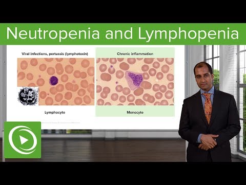
Viral Infections and Lymphocytosis
- Viral infections can lead to lymphocytosis; Bordetella pertussis inhibits lymphocyte entry into lymph nodes causing absolute lymphocytosis.
Chronic Inflammation and Monocytosis
- Monocytosis often indicates chronic inflammation where neutrophils are replaced by monocytes after their apoptosis around day three post-infection.
Neutrophilic Leukocytosis
- Neutrophilic leukocytosis occurs when there’s an increase in neutrophil counts above the normal range of 2,200 - 5,600 cells per microliter.
- This condition is commonly associated with acute inflammation or bacterial infections.
Neutrophilic Leukocytosis and Its Implications
Neutrophil Dynamics in Inflammation
- Neutrophils play a crucial role in the inflammatory response, particularly after an infarction. Around the third day post-infarction, neutrophils undergo apoptosis and are replaced by monocytes or macrophages.
- Corticosteroids are identified as common drugs that cause neutrophilic leukocytosis through d-margination, which inhibits adhesion molecules leading to decreased margination of neutrophils and an increase in circulating neutrophil pools.
Cytokine Influence on Bone Marrow Activity
- Interleukin 17 is highlighted as a significant cytokine that stimulates bone marrow activity during acute inflammation, resulting in increased production of neutrophils.
- During heightened bone marrow activity, immature forms of neutrophils (band cells) may also be released into circulation alongside mature segmented neutrophils.
Understanding Left Shift in Pathology
- The term "left shift" refers to the presence of more immature band cells in circulation during episodes of neutrophilic leukocytosis, indicating increased recruitment from the bone marrow.
- A left shift signifies a higher number of band cells due to acute inflammation; this contrasts with normal maturation processes where cells progress from primitive (left) to mature (right).
Clinical Cases: Appendicitis and Sepsis
- An extreme case of neutrophilic leukocytosis can occur with conditions like acute myocardial infarction or appendicitis, where WBC counts exceed 7000.
- In young children presenting with lymphoid hyperplasia and right lower quadrant pain, appendicitis is a primary differential diagnosis.
Complications During Pregnancy
- Pregnant women face increased susceptibility to urinary tract infections due to anatomical changes; this can escalate to severe conditions such as sepsis if not managed properly.
- Severe cases like perforated appendicitis or sepsis can lead to WBC counts exceeding 50,000, resembling leukemia but termed "leukemoid reaction" because these elevated counts consist of functional neutrophils.
Differentiating Leukemoid Reaction from Leukemia
- Unlike leukemia where dysfunctional cells predominate, leukemoid reactions involve functional neutrophils responding effectively to infection.
- Laboratory tests assessing functionality include alkaline phosphatase levels; high levels indicate active recruitment during infections compared to low levels seen in chronic myelogenous leukemia (CML).
Molecular Mechanisms in Pathology
- The discussion transitions into molecular pathology involving the JAK2/STAT pathway. Abnormal findings such as fibrosis within the bone marrow suggest underlying pathologies affecting hematopoiesis.
Bone Marrow Infiltration and Its Implications
Understanding Bone Marrow Lesions
- The discussion begins with the potential impact of breast cancer on bone marrow, highlighting how invasive lobular breast cancer can lead to metastasis in the bone marrow, creating space-occupying lesions that displace normal stem cells.
- The presence of various cell types in the bone marrow, such as megakaryocytes and nucleated red blood cells (RBCs), is noted. These cells are pushed out due to the space-occupying lesions caused by metastatic cancer.
Consequences of Displacement
- When normal hematopoietic cells are displaced into circulation due to these lesions, it results in a phenomenon known as leukemoid reaction characterized by an increase in immature white blood cells (WBCs) and RBC blasts.
- The significance of finding nucleated RBCs in peripheral blood smears is emphasized; their presence indicates abnormal conditions since they should not typically be found outside the bone marrow.
Neutrophilic Leukocytosis Explained
- An extreme case of neutrophilic leukocytosis may arise from infiltrative diseases or multiple fractures affecting the bone marrow, leading to increased immature WBC release into circulation.
- The commonality between cases involving breast cancer metastasis and multiple fractures is identified: both scenarios result in infiltration of the bone marrow, causing similar hematological responses.
Distinguishing Features from Leukemia
- A comparison is made between this condition and leukemia; while there may be an increase in blasts, patients do not exhibit typical leukemic symptoms like anemia or thrombocytopenia.
- It’s clarified that only certain cell types like bands might be present without indicating full-blown leukemia symptoms despite elevated blast counts.
Peripheral Blood Smear Analysis
- Observations from peripheral blood smear images reveal tear drop-shaped RBCs and nucleated RBCs indicative of underlying issues such as infiltration due to fibrosis or metastases from cancers like breast cancer.
- The metaphorical use of "tear drop" for affected RBC shapes illustrates the distressing impact on normal erythropoiesis within the context of space occupying lesions.