Testículo y vías masculinas - Reproductor y desarrollo
Introduction to Male Genital Anatomy
Overview of the Male Reproductive System
- The male genital system consists of internal and external organs, including testicles, epididymis, vas deferens, urethra, and accessory sexual glands such as seminal vesicles and prostate.
- External genitalia include the penis and scrotum. The testicles are located in the scrotum outside the abdominal cavity.
Structure and Function of Testicles
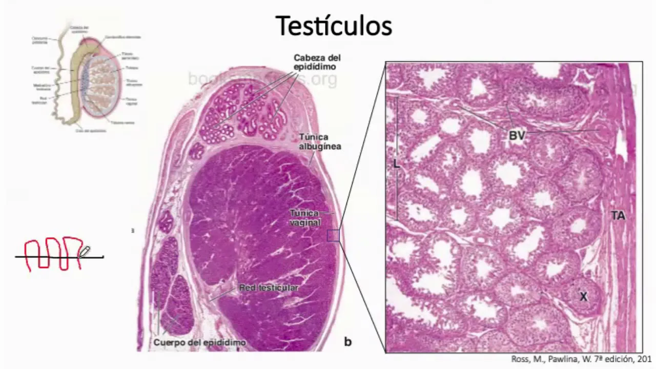
- Testicles are oval-shaped, measuring approximately 2-3 cm wide and 4 cm high; their primary functions include gamete formation (spermatogenesis) and hormone secretion (testosterone).
- Each testicle is covered by a dense connective tissue capsule called tunica albuginea, which sends septa into the organ to create lobules containing seminiferous tubules.
Seminiferous Tubules and Sperm Production
Anatomy of Seminiferous Tubules
- Each lobule contains about 2 to 4 seminiferous tubules that can be up to 50 cm long when uncoiled; they are highly contorted to fit within the testicle's dimensions.
- The seminiferous tubules connect to straight tubules that lead into a network known as rete testis located at the mediastinum of the testicle.
Pathway for Sperm Maturation
- Rete testis communicates with efferent ducts leading to the epididymis where sperm maturation occurs.
Developmental Aspects of Testicular Positioning
Descent of Testes
- During fetal development, testes form in the abdominal cavity before descending into the scrotum; this positioning is crucial for spermatogenesis which requires lower temperatures than body temperature.
Implications of Undescended Testes
- If testes do not descend properly (cryptorchidism), it can lead to infertility due to elevated temperatures affecting sperm production.
Histological Structure of Testes
Layers Surrounding Testes
- The tunica vaginalis covers each testicle with two layers: visceral (adhering directly to tunica albuginea) and parietal layers.
Vascularization and Tissue Composition
- Tunica albuginea is composed of dense connective tissue with blood vessels supplying nutrients; it divides into lobules containing seminiferous tubules interspersed with Leydig cells responsible for testosterone synthesis.
Role of Leydig Cells in Hormone Production
Functionality within Interstitial Space
- Leydig cells located between seminiferous tubules synthesize testosterone which enters circulation while spermatogenesis occurs within those tubules.
Histological Analysis of Testicular Structure
Overview of Leydig Cells and Steroid Production
- The transcript begins with a description of testicular tissue, highlighting the presence of blood vessels and Leydig cells, which are steroid-producing cells.
- It is noted that steroids are synthesized from cholesterol, with mitochondria exhibiting tubular characteristics typical for steroid-producing organs.
- Leydig cells produce testosterone during fetal development, crucial for the formation of male genital organs. Their activity ceases around 55 days into fetal life.
Developmental Changes in Leydig Cells
- After initial testosterone production, Leydig cells retract but remain present until puberty when they re-develop under the influence of pituitary hormones.
- Testosterone plays a vital role in initiating spermatogenesis and developing secondary male sexual characteristics during puberty.
Structure and Function of Seminiferous Tubules
- The seminiferous tubules contain stratified epithelium composed of various cell types at different heights; not all contact the basal membrane.
- Spermatogenic cells are responsible for producing spermatozoa; these include spermatogonia in contact with the basal membrane.
Stages of Spermatogenesis
- Different stages within spermatogenesis are identified, including differentiating spermatocytes transitioning towards becoming mature spermatozoa.
- Sertoli cells support spermatogenic processes by contacting the basal membrane and facilitating sperm movement through contraction.
Detailed Process of Spermatogenesis
- Spermatogenesis involves several phases: starting from spermatogonia undergoing mitosis to form syncytia that synchronize differentiation.
- The process includes transitions through various types (A dark, A light, B), where mitotic divisions lead to interconnected cellular structures sharing cytoplasmic components.
Meiosis and Sperm Maturation
- In meiosis, diploid cells undergo two consecutive divisions to become haploid spermatozoa. This transformation is critical for successful reproduction.
- Following meiosis, sperm undergo morphological changes such as chromatin condensation and flagellum development while losing excess cytoplasm.
Final Stages Before Sperm Release
- Mature sperm detach from Sertoli cells into the lumen of seminiferous tubules after completing their transformation process known as spermiogenesis.
- Residual bodies formed during this process are phagocytized by Sertoli cells, ensuring efficient use of resources within the testicular environment.
Understanding Spermatogenesis and Cellular Associations
Overview of Spermatogonias and Their Differentiation
- Spermatogonias are cells in contact with the basal lamina of the epithelium, transitioning to different stages of sperm development as they move towards the lumen.
- The early spermatogenic cells progress through various differentiation stages, forming distinct cellular associations within the seminiferous tubules.
Cellular Combinations and Cycles
- In humans, there are six possible combinations of cell types at different differentiation stages throughout the seminiferous tubules.
- These combinations reflect a cycle known as "the seminiferous epithelium cycle," which describes how long it takes for a specific cellular association to reappear in a given region.
Duration of Spermatogenesis
- The complete process of spermatogenesis takes approximately 74 days, involving multiple cycles (46 cycles noted).
- The distribution patterns of these cellular associations vary between species; in mice, sequential distributions occur while humans exhibit patchy distributions across cross-sections.
Role of Leydig Cells and Hormonal Production
- Leydig cells produce testosterone during fetal development but cease until puberty when they resume production, essential for spermatogenesis.
- Prepubescent males show immature seminiferous tubules with only spermatogonia present; adult males have more complex structures with various cell types.
Sertoli Cells Functionality
- Sertoli cells support and nourish developing sperm cells; they also phagocytize residual cytoplasm from maturing spermatozoa.
- These cells form the blood-testis barrier and synthesize proteins necessary for androgen binding, crucial for testosterone function during development.
Characteristics of Sertoli Cells
Complex Cellular Junctions in Spermatogenesis
Characteristics of Sertoli Cell Junctions
- The transcript discusses a specific type of cellular junction known as the Sertoli cell junction, highlighting its unique structural characteristics.
- These junctions are described as tight and dense, featuring over 50 fusion lines that contribute to their hermetic nature.
- Actin filaments play a crucial role in maintaining the integrity of these junctions, interspersed between the membranes of adjacent cells.
Compartments Formed by Tight Junctions
- The tight junctions between Sertoli cells create two distinct compartments: a basal compartment for spermatogonia and an adluminal compartment for differentiating spermatozoa.
- The composition of the adluminal compartment is similar to blood plasma, containing ions, amino acids, carbohydrates, and proteins essential for sperm development.
Hormonal Influence and Antigen Presentation
- The basal compartment has a different ionic and protein composition with high testosterone levels transported by androgen-binding protein synthesized by Sertoli cells.
- It is noted that these cells present antigens distinct from those of the individual’s own tissues, which is significant for immune tolerance during spermatogenesis.
Structure and Function of Seminiferous Tubules
- The seminiferous tubules lead into straight tubules lined only by Sertoli cells before connecting to the rete testis located in the mediastinum testis.
- These structures are covered with simple cuboidal epithelium and have minimal microvilli; they serve as conduits for sperm transport.
Epididymis Structure and Sperm Maturation
- The epididymis measures approximately 7.5 cm long and consists of coiled ducts where sperm maturation occurs.
- Ductal epithelium features tall columnar ciliated cells that help move sperm from seminiferous tubules towards the epididymis.
Role of Smooth Muscle in Sperm Transport
- Surrounding smooth muscle layers contract to facilitate sperm movement through the reproductive tract toward ejaculation.
- The appearance of ductal lumens is influenced by epithelial cell types present—ciliated columnar and cuboidal with microvilli aiding reabsorption processes.
Storage and Capacitation of Spermatozoa
- In the epididymal lumen, sperm are stored until ejaculation; this environment allows them to gain motility while acquiring surface factors necessary for fertilization.
- A reversible inhibitory factor on sperm capacitation is discussed; it detaches upon reaching female reproductive tracts allowing successful fertilization.
Epithelial Composition in Reproductive Tract
- Throughout most male reproductive pathways, stratified epithelium composed of tall columnar principal cells with stereocilia participates in reabsorbing luminal elements.
Overview of Male Reproductive Anatomy
Changes in Smooth Muscle Cells
- The layers of smooth muscle cells will increase in proportion, while cylindrical cells will decrease in height; at the head, they measure approximately 80 micrometers, and at the tail, about 40 micrometers.
Epididymis and Ductus Deferens
- A comparison between the epididymis and ductus deferens shows that the latter has a more pronounced lumen, while the epididymis is characterized by its taller principal cells.
- The ductus deferens is the longest male genital tract, connecting the epididymis to the ejaculatory ducts, measuring around 40 centimeters. It features a mucosa made up of pseudostratified columnar epithelium with basal and principal cells.
Structure of Ductus Deferens
- The ductus deferens is surrounded by several muscular layers: an inner longitudinal layer where cells are arranged longitudinally towards the lumen and an outer circular layer.
- Contraction of these muscular layers occurs via sympathetic stimulation during ejaculation. Accessory sexual glands include seminal vesicles, prostate gland, and bulbourethral glands.
Seminal Vesicles Functionality
- Seminal vesicles are tubular organs that secrete a thick yellowish component constituting about 70% of semen; this secretion contains proteins and coagulating factors.
- The mucosa of seminal vesicles features folds lined with pseudostratified columnar epithelium with secretory cells.
Prostate Gland Characteristics
- The prostate surrounds the first portion of the urethra; it secretes a milky fluid that constitutes about 30% of ejaculate volume.
- Prostatic secretion has a slightly acidic pH due to citric acid content and includes prostate-specific antigen (PSA), which prevents coagulation.
Histological Features of Prostate
- The prostate consists of tubuloalveolar glands lined with simple or pseudostratified columnar epithelium.
- Amylaceous bodies may be found within glandular lumina as age progresses; excretory ducts lead into the urethra.
Regions Within Prostate Gland
- The prostate can be divided into three zones: peripheral (containing main glands), central (with mucous glands), and transitional regions releasing secretions into urethra.
Bulbourethral Glands Role
Overview of Male Reproductive Anatomy
Glands and Their Functions
- The male reproductive system includes tubular glands, specifically the seminal vesicles, prostate gland, and bulbourethral glands. These structures release secretions into the urethra.
Structure of the Penis
- The penis consists of three cylindrical bodies: two corpora cavernosa and one corpus spongiosum, which contains the urethra.
Tissue Composition
- Each erectile body is surrounded by a dense connective tissue layer called the tunica albuginea. This structure provides support and maintains shape during erection.
Skin and External Features
- The outermost layer of the penis is skin, with a specific mention of the glans (the tip).
Erectile Tissue Mechanics
- The erectile tissue in both corpora cavernosa and corpus spongiosum comprises blood-filled spaces that expand during arousal due to increased blood flow.
Muscle Functionality During Erection
- Smooth muscle contractions regulate blood flow; parasympathetic stimulation leads to relaxation and filling of blood spaces, causing an erection.
Urethra Segmentation
- The urethra measures approximately 20 cm long with four segments: pre-prostatic (transitional epithelium), prostatic (also transitional), membranous (pseudostratified columnar epithelium), and penile urethra.
Associated Glands