🥇 Anatomía del PÁNCREAS y los CONDUCTOS PANCREÁTICOS. Fácil, Rápido y Sencillo
Introduction to the Anatomy of the Pancreas
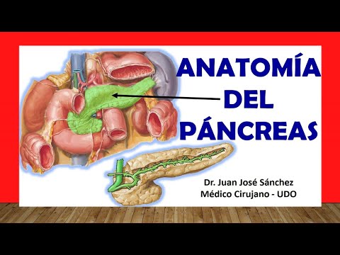
In this video, Dr. Juan José Sánchez provides an overview of the anatomy of the pancreas. He discusses its importance as a vital organ and its role in both endocrine and exocrine functions.
Generalities about the Pancreas
- The pancreas is a retroperitoneal organ, meaning it is located behind the peritoneum.
- It is a mixed gland, producing both hormones (endocrine function) and pancreatic juices (exocrine function).
- The pancreas has three main portions: head, body, and tail.
- The head of the pancreas is connected to the concavity of the duodenum.
- The body of the pancreas is elongated and connects to the head at a narrow portion called the neck.
- The tail of the pancreas is distal and closely related to the spleen through the splenorenal ligament.
Relationships and Structures
- The posterior surface of the stomach is closely related to the anterior surface of the pancreas.
- Between these two structures lies a space known as "transcavidad de los epiplones" or greater omental bursa.
- The uncinate process is a hook-shaped extension located at the junction between the head and body of the pancreas.
- It extends towards left and upward behind superior mesenteric vessels.
- This process plays an important role in anatomical relationships with adjacent structures such as superior mesenteric vein.
- The pancreatic ducts terminate in the head of the pancreas, including common bile duct which perforates through it.
Divisions and Parts of Pancreas
Dr. Juan José Sánchez explains in detail about different parts and divisions of the pancreas.
Divisions of the Pancreas
- The pancreas has three main portions: head, body, and tail.
- The junction between the head and body is called the neck.
- The tail is the most distal part of the pancreas.
Tubérculo Pancreático (Pancreatic Tubercle)
- Located in the superior anterior part of the pancreas.
- It is an elevation just below the celiac trunk.
Ligamento Esplénico Renal (Splenic Renal Ligament)
- Connects the tail of the pancreas to the spleen.
- Also connects to the kidney.
Anatomical Relationships
Dr. Juan José Sánchez discusses important anatomical relationships involving the pancreas.
Relationship with Stomach and Transcavidad de los Epiplones
- The posterior surface of the stomach is closely related to the anterior surface of the pancreas.
- Between them lies a space known as "transcavidad de los epiplones" or greater omental bursa.
Relationship with Duodenum
- The head of the pancreas is directly connected to the concavity of duodenum.
- Secretions from pancreatic ducts enter into second portion of duodenum for digestion purposes.
Conclusion
Anatomy of the Pancreas
This section provides an overview of the anatomical relationships of the pancreas, including its connections to other organs and structures within the abdominal cavity.
Relationships of the Pancreas
- The transverse mesocolon supports the transverse colon and passes anteriorly through the pancreas.
- The posterior surface of the stomach is closely related to the anterior surface of the pancreas, forming the omental bursa or lesser sac.
- Posterior to the head of the pancreas are the inferior vena cava, abdominal aorta, and common bile duct.
- The right kidney, renal artery, renal vein, and gonadal artery are also posterior relations of the pancreatic head.
- The diaphragm and superior mesenteric artery lie posteriorly to the body of the pancreas.
- The left adrenal gland and left kidney are related to the body and tail of the pancreas.
- The splenic vein and artery are closely associated with both body and tail regions.
Pancreatic Ducts
This section discusses important ducts associated with pancreatic function.
Pancreatic Ducts
- The common bile duct carries bile from liver to duodenum. It joins with Wirsung's duct (main pancreatic duct) at a dilated region called ampulla of Vater or hepatopancreatic ampulla.
- Wirsung's duct extends from tail to head of pancreas. It unites with common bile duct in second part of duodenum for secretion into digestive system.
- Santorini's duct is an accessory pancreatic duct that drains a portion of pancreatic tissue into duodenum. Not everyone has this accessory duct.
Sphincters and Papillae
This section explains the sphincters and papillae associated with the pancreatic ducts.
Sphincters and Papillae
- The Oddi's sphincter is located at the junction of the common bile duct and duodenum. It controls the flow of bile and pancreatic secretions into the duodenum.
- The pancreatic sphincter regulates the release of pancreatic secretions through Santorini's duct.
- The major duodenal papilla (caruncula) is a larger opening where both common bile duct and main pancreatic duct empty their contents.
- The minor duodenal papilla (caruncula) is a smaller opening specifically for Santorini's duct to secrete its product.
Anatomy of the Pancreas
This section provides an overview of the anatomy of the pancreas, including the sphincter of the ampulla of Vater and the pancreatic sphincter.
Sphincters of the Ampulla of Vater
- The blue-colored sphincter is known as the hepatopancreatic sphincter or sphincter of Oddi.
- The pancreatic sphincter is located within the ampulla of Vater.
Irrigation of the Pancreas
This section discusses the blood supply to the pancreas and its importance due to its endocrine function.
Blood Supply to the Head of Pancreas
- The head of the pancreas is supplied by two major arteries:
- Anterior pancreaticoduodenal artery (branching from gastroduodenal artery)
- Posterior pancreaticoduodenal artery (located behind the head)
Formation of Pancreaticoduodenal Arches
This section explains how pancreaticoduodenal arches are formed by specific arteries.
- The anterior pancreaticoduodenal arch is formed by:
- Anterosuperior pancreaticoduodenal artery (direct branch from gastroduodenal artery)
- Anteroinferior pancreaticoduodenal artery (branching from inferior pancreaticoduodenal trunk)
Blood Supply to Body and Tail of Pancreas
This section describes how blood supply reaches the body and tail regions of the pancreas.
- The posterior pancreaticoduodenal arch is formed by:
- Posteroinferior pancreaticoduodenal artery (branching from inferior pancreaticoduodenal trunk)
- Pancreaticoduodenal artery (superior branch of gastroduodenal artery)
Venous Drainage of the Pancreas
This section explains the venous drainage system of the pancreas.
- Venous drainage is primarily through pancreaticoduodenal veins, which form anterior and posterior arches.
- These veins drain into the portal vein and superior mesenteric vein.
- The splenic vein also contributes to venous drainage.
Innervation of the Pancreas
This section discusses the innervation of the pancreas by both sympathetic and parasympathetic systems.
- Sympathetic innervation is provided by ganglia in the celiac plexus and superior mesenteric plexus.