Fibrilación ventricular y taquicardia ventricular sin pulso
Fibrilación Ventricular y Taquicardia Ventricular Sin Pulso
Introducción a la Fibrilación Ventricular y Taquicardia Ventricular Sin Pulso
- Fernanda de Cirema Maldonado Marroquín y José Andrés de León Solares presentan una clase sobre fibrilación ventricular y taquicardia ventricular sin pulso, temas previamente revisados en documentos y diapositivas.
Definición y Características
- La fibrilación ventricular (FV) y la taquicardia ventricular sin pulso (TVSP) son arritmias letales que causan paro cardíaco súbito al impedir la contracción efectiva del corazón, afectando el flujo sanguíneo.
- FV se caracteriza por actividad eléctrica caótica, mientras que TVSP presenta actividad ordenada pero sin pulso palpable. Ambas requieren reconocimiento inmediato, RCP de alta calidad y desfibrilación temprana.
Importancia Clínica
- Según las guías de la American Heart Association 2023, la fibrilación ventricular es una causa frecuente de muerte súbita, constituyendo una emergencia médica que demanda intervención inmediata.
- En casos de paro cardíaco súbito, hay un 90% de probabilidad que sea debido a FV o TVSP.
Diagnóstico Electrocardiográfico
- Los valores ECG para FV incluyen complejos QRS indeterminados con ausencia de ondas P y T reconocibles; frecuencias entre 150 a 500 por minuto indican ritmo caótico.
- Se muestra un algoritmo utilizado por la American Heart Association para el manejo inicial en caso de FV o TVSP.
Manejo Inicial en Emergencias
- En un paciente en paro cardíaco, se inicia RCP inmediatamente; se conecta al monitor para determinar si el ritmo es desfibrilable.
- Si el ritmo es desfibrilable (FV o TVSP), se realiza la primera descarga después de dos minutos de RCP. Se debe establecer acceso intravenoso o intraóseo si no es posible.
Protocolo Avanzado
- Después del segundo ciclo de RCP, se administra adrenalina cada 3 a 5 minutos; considerar intubación si es necesario.
- Se administran medicamentos como amiodarona o lidocaína tras confirmar nuevamente si el ritmo sigue siendo desfibrilable.
Causas Reversibles
- Es crucial identificar causas reversibles durante el tratamiento: hipovolemia, hipoxia, acidosis metabólica (hidrogeniones), desequilibrios electrolíticos (hipo/hiperpotasemia), hipotermia, tensión neumotóxica, etc.
Detalles sobre Taquicardia Ventricular Sin Pulso
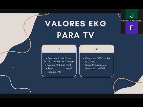
- La TVSP tiene frecuencia cardíaca mayor a 100 latidos por minuto con origen bajo el haz de His; los complejos QRS son anchos (>0.12 segundos).
Resumen Final sobre Algoritmos
- El mismo algoritmo aplicado para FV también se utiliza para TVSP; ambos requieren atención rápida e intervención adecuada para restaurar ritmos efectivos.
Clinical Management of Ventricular Tachycardia
Case Presentation
- A clinical case is introduced involving a 58-year-old male patient with a history of hypertension, who collapsed at home and was brought to the hospital unconscious.
- Upon arrival, the patient was found to be unconscious with carotid pulse and spontaneous respiration, raising suspicion for a defibrillable rhythm due to sudden loss of consciousness.
Initial Assessment and Monitoring
- The first step in management involves connecting the patient to a monitor or defibrillator to assess the cardiac rhythm.
- The monitor displayed a chaotic irregular rhythm without identifiable QRS complexes, indicating possible ventricular fibrillation.
CPR Protocol
- Recognition of cardiac arrest is crucial; safety measures are taken before initiating CPR. High-quality CPR begins after confirming absence of pulse and breathing.
- High-quality CPR characteristics include compressions at 100-120 per minute, depth of 5-6 cm, complete chest recoil, and changing rescuers every 2 minutes.
Defibrillation Process
- The patient was ventilated using bag-valve-mask at a ratio of 30 compressions to 2 breaths since no advanced airway was available.
- After identifying a defibrillable rhythm (ventricular fibrillation), the first shock was delivered. Both ventricular fibrillation and pulseless ventricular tachycardia follow similar management protocols.
Shock Administration Guidelines
- Defibrillators can be biphasic or monophasic; biphasic is more common in hospitals. Recommended energy levels are typically 120–200 J for biphasic and up to 360 J for monophasic shocks.
- Post-defibrillation, CPR should continue for another two minutes without checking pulses until reevaluation occurs.
Medication Administration
- If the patient remains in a defibrillable rhythm after two minutes, a second shock is administered followed by epinephrine (1 mg IV/IO every 3–5 minutes).
- Epinephrine administration includes elevating the patient's arm for about ten seconds followed by flushing with saline (10 cc).
Continued Resuscitation Efforts
- After administering epinephrine, another two-minute cycle of CPR follows before reassessing the patient's rhythm.
- If still in ventricular fibrillation or pulseless VT after three shocks, amiodarone (300 mg IV/IO bolus), may be given with an additional dose if necessary based on subsequent rhythms.
Cardiac Arrest Management and Post-Resuscitation Care
Key Steps in Cardiac Arrest Management
- Always alternate compressions, defibrillation, and medication while evaluating the rhythm every 2 minutes. Check for reversible causes using the "5 Hs" and "5 Ts" to understand why the patient remains in cardiac arrest despite interventions.
- The "5 Hs" include:
- Hypoxia
- Hypovolemia
- Hydrogen ions (acidosis)
- Hypo/hyperkalemia
- Hypothermia
- The "5 Ts" consist of:
- Tamponade
- Tension pneumothorax
- Toxins
- Thrombosis (coronary)
The goal is to achieve return of spontaneous circulation (ROSC).
Post-Arrest Management
- If ROSC is achieved, stabilize the airway and ensure oxygen saturation is above 94%. Manage blood pressure carefully due to potential ischemic periods experienced by the patient. Monitor for signs of myocardial infarction and consider urgent coronary angiography if necessary.
- Maintain controlled temperature, monitor blood glucose levels, and provide organ support as needed during post-arrest care. This includes assessing for any complications that may arise after resuscitation efforts.
Understanding Tachyarrhythmias
- Tachyarrhythmia is characterized by a heart rate exceeding 150 beats per minute but does not equate to cardiac arrest since there is still a pulse present. Symptoms can include hypotension, altered mental status, shock signs, chest pain, or acute heart failure.
- Differentiate between typical rhythms and tachyarrhythmias; irregular impulses can lead to tachycardia requiring specific management protocols rather than standard CPR techniques used in cardiac arrest situations.
Initial Assessment and Treatment of Tachyarrhythmias
- For patients with a heart rate ≥150 bpm exhibiting hemodynamic compromise symptoms, immediate assessment of airway patency and oxygenation is crucial along with continuous cardiac monitoring to identify rhythm type. Control blood pressure and oximetry throughout treatment processes.
- If no significant symptoms are present but QRS duration exceeds 12 seconds, establish intravenous access for potential antiarrhythmic infusion or consult an expert if available for further management strategies tailored to the patient's condition.