PLEXO BRAQUIAL - PARTE 1
Overview of the Brachial Plexus
Introduction to the Brachial Plexus
- The brachial plexus is discussed as a crucial network formed by cervical nerves, particularly focusing on its roots and their anatomical significance.
- The presentation highlights both the supraclavicular and infraclavicular parts of the brachial plexus, emphasizing their roles in upper limb innervation.
Key Nerves Originating from the Brachial Plexus
- Important nerves originating from the brachial plexus include: musculocutaneous nerve, axillary nerve, median nerve, radial nerve, and ulnar nerve.
- These nerves are essential for examining upper limb functionality during clinical assessments.
Anatomical Relationships
- The relationship between the brachial plexus and surrounding muscles (specifically scalene muscles) is outlined, indicating how these structures interact anatomically.
- The connection between the brachial plexus and subclavian artery is also noted, highlighting its relevance in understanding vascular relationships.
Spinal Anatomy Related to Brachial Plexus
Vertebral Structure and Meninges
- A detailed description of vertebral anatomy includes components like vertebral body, arch, spinous processes, and transverse processes.
- Discussion on spinal meninges (dura mater, arachnoid mater, pia mater), emphasizing their protective role around neural tissue.
Nerve Roots and Their Pathways
- Explanation of how spinal nerves traverse intervertebral foramina with branches forming peripheral nerves; this includes dorsal (posterior) and ventral (anterior) rami.
Components of the Brachial Plexus
Formation of the Brachial Plexus
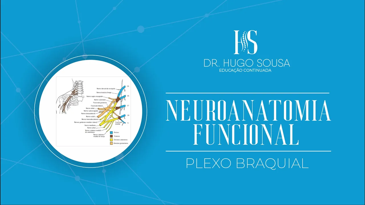
- The brachial plexus consists primarily of roots C5 to T1; contributions may also come from C4 in some cases.
Trunks of the Brachial Plexus
- Three main trunks form from these roots: superior trunk (C5-C6), middle trunk (C7), and inferior trunk (C8-T1).
Divisions within the Brachial Plexus
Anterior and Posterior Divisions
- Two divisions exist within each trunk: anterior division which typically supplies flexor muscles; posterior division which supplies extensor muscles.
Plexus Brachialis Overview
Structure of the Plexus Brachialis
- The anterior division of the plexus is formed by the superior trunk and a union with the middle trunk, which does not have a superior component. This combination creates the anterior division alongside elements from all three trunks: superior, middle, and inferior.
- The posterior division receives contributions from all three trunks (superior, middle, and inferior), indicating a comprehensive integration within the brachial plexus structure.
Fascicles of the Plexus Brachialis
- There are three main fascicles in the brachial plexus: lateral, medial, and posterior. The lateral fascicle is formed from fibers originating from both the superior and middle trunks.
- The medial fascicle arises from roots C8 and T1. In contrast, the posterior fascicle integrates information from all three trunks (superior, middle, and inferior).
Terminal Branches of the Plexus Brachialis
- From these fascicles emerge terminal branches that include several key nerves: musculocutaneous (C5-C7), axillary (C5-C6), radial (C5-T1), median (C5-T1), and ulnar (C7-T1).
- The radial nerve is noted as being particularly thick among these nerves; it encompasses roots ranging from C5 to T1. The median nerve has both lateral and medial divisions contributing to its formation.
Additional Nerves Emerging from Roots
- Some significant nerves also emerge directly from root portions of the brachial plexus. For instance, notable contributions include dorsal scapular nerve for muscle innervation.
- Other important nerves such as phrenic nerve arise here as well; they play crucial roles in diaphragm function.
Innervation Functions
- Specific muscles like scalenes receive innervation through these emerging nerves; they are essential for neck flexion movements.
- Within trunk regions of the plexus, additional nerves like suprascapular nerve contribute to shoulder muscle innervation.
Understanding the Brachial Plexus and Associated Pathologies
Overview of the Brachial Plexus
- The lecture begins with a discussion on the brachial plexus, focusing on its anterior and posterior divisions, as well as terminal branches.
- The relationship between the subclavian artery and axillary artery is highlighted, along with the organization of trunks within the plexus: superior, medial, and inferior trunks.
Terminal Branches and Clinical Relevance
- The importance of terminal branches originating from fascicles in relation to clinical evaluations is emphasized.
- Various conditions related to brachial plexus injuries are introduced, including obstetric paralysis due to traumatic avulsions.
Types of Obstetric Paralysis
- Two types of obstetric paralysis are discussed: high paralysis (Erb's palsy), typically involving C5-C6 roots, and lower paralysis (Klumpke's paralysis), associated with C7-C8-T1 roots.
- Klumpke's paralysis is characterized by its impact on lower brachial plexus function.
Causes of Brachial Plexus Disorders
- Other causes for brachial plexopathy include tumors that disrupt innervation and complications from radiotherapy leading to fibrosis.
- Traumatic avulsions from accidents, particularly motorcycle incidents where excessive stretching occurs during falls, are noted as common causes.
Observations in Pediatric Cases
- Obstetric-related avulsions can occur during childbirth; this risk is higher in hypotonic infants.
- Complications arising from delivery mechanisms can lead to significant functional impairments in children.
Characteristic Hand Deformities
- Specific hand deformities resulting from nerve injuries are described:
- "Claw hand" due to ulnar nerve injury.
- "Wrist drop" linked to radial nerve damage.
- "Benediction hand" associated with median nerve issues.
Testing Integrity of the Brachial Plexus
- Methods for assessing brachial plexus integrity are crucial for understanding motor function in upper limbs.
- The influence of specific root levels (C5-T1) on shoulder movements such as abduction/adduction and rotation is outlined.
Functional Movements Related to Nerve Roots
- Detailed connections between spinal roots and their corresponding muscle functions are provided:
- Flexion/extension at elbow joint involves C5-C6 for flexion and C7 for extension.
- Pronation/supination movements rely on C6-C8 roots.
Understanding Upper Limb Movements and Nerve Involvement
Overview of Joint Movements and Spinal Segments
- The discussion begins with an overview of joint movements and the spinal segments involved, emphasizing the importance of testing root integrity through movement.
Nerves Associated with Shoulder Abduction
- For shoulder abduction, the suprascapular nerve is highlighted as a key player in this movement, alongside other nerves that contribute to shoulder function.
Nerves Related to Adduction and Rotation
- Adduction involves both the thoracodorsal nerve and medial/lateral pectoral nerves. The conversation also touches on lateral rotation movements influenced by various nerves including the suprascapular nerve.
Elbow Flexion and Extension Mechanics
- The musculocutaneous nerve is identified as responsible for elbow flexion, while extension is governed by the radial nerve. This section underscores how specific nerves control distinct movements at the elbow joint.
Hand Movement Control via Nerves