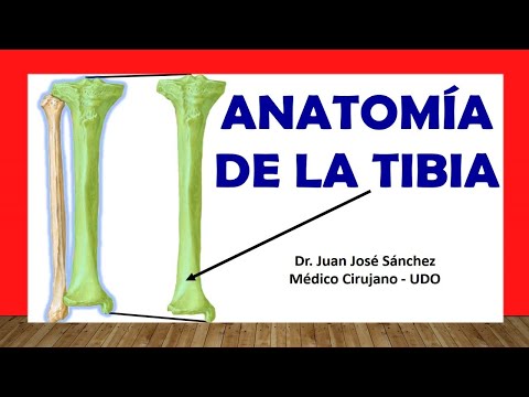
🥇 Anatomía de La TIBIA, Fácil, Rápida y Sencilla
Anatomy of the Tibia
In this video, the speaker discusses the anatomy of the tibia, highlighting its structure and key features within the lower limb.
Divisions of Lower Limb
- The lower limb is divided into four portions: pelvic girdle (coxal bone), thigh region (femur), leg region (tibia and fibula), and foot (tarsus, metatarsals, phalanges).
Osteology of Tibia
- The tibia is classified as a long bone with two epiphyses (proximal and distal) and a diaphysis.
- Detailed breakdown: proximal epiphysis, diaphysis, and distal epiphysis.
Upper Epiphysis Features
- Tibia's position relative to fibula; medial to fibula in the leg.
- Identification of condyles - lateral and medial; articulation with femur's condyles.
Intercondylar Zones
- Description of intercondylar zones between condyles; anterior and posterior regions.
- Explanation of intercondylar eminence dividing anterior and posterior areas.
Diaphysis Structure
- Origin points for cruciate ligaments on tibia.
Anatomy of the Tibia
In this section, the anatomy of the tibia is discussed in detail, focusing on its edges, faces, and specific features.
The Edges and Faces of the Tibia
- The tibia has three main edges: anterior, medial/internal, and lateral/external.
- These edges form different faces of the tibia: posterior face between medial and lateral edges, along with lateral, medial, and anterior faces.
- The posterior edge of the tibia presents anatomical features like the intercondylar area with an oblique line and a nutritional foramen for artery supply.
Lower Epiphysis and Medial Malleolus
- The lower epiphysis of the tibia includes the internal malleolus or medial malleolus with five distinct faces: anterior, lateral, posterior, medial, and inferior.
- Notable features include the malleolar groove on the posterior face for tendons attachment and a notch for fibular articulation towards laterally.
Articulation Points and Functionality
- Two tibiofibular joints are formed by notches on the internal malleolus to articulate with the fibula at proximal and distal points.