PLEXO CERVICAL - PARTE 2
Plexo Cervical: Estrutura e Funções
Introdução ao Plexo Cervical
- O plexo cervical está localizado na região do pescoço, formado pelas vértebras cervicais. Os nervos que o compõem se originam da região cervical.
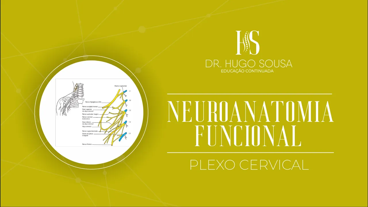
- Quatro ramos dos nervos espinhais cervicais (C1, C2, C3 e C4) formam o plexo cervical. As raízes de C1 a C4 são essenciais para sua formação.
Conexões Nervosas
- As raízes dos nervos estabelecem conexões para inervar regiões específicas do corpo. Essas conexões são fundamentais para a função motora e sensitiva.
- O nervo hipoglosso (12º par craniano), responsável pela motricidade da língua, conecta-se com as raízes do plexo cervical, destacando a inter-relação entre os sistemas nervosos.
Alça Cervical
- A alça cervical é formada por contribuições das raízes de C1, C2 e C3. Essa estrutura é crucial para a inervação de músculos no pescoço.
- A alça possui uma raiz superior (C1) e uma raiz inferior (C2 e C3), sendo importante para a coordenação muscular na região cervical.
Ramos Nervosos
- Existe conexão entre as raízes de C3 e C4 que se unem para formar importantes nervos como o occipital menor e auricular magno.
- O nervo frênico, que também recebe contribuições de C5, é vital para a inervação do diafragma, mostrando como as raízes cervicais influenciam funções respiratórias.
Territórios de Inervação
- Os ramos cutâneos superficiais do plexo cervical incluem o nervo occipital menor (inerva couro cabeludo), auricular magno (inerva pele ao redor da orelha), e outros que afetam áreas superiores ao tórax.
- Os ramos motores profundos estão relacionados à alça cervical e ao nervo frênico, inervando músculos profundos do pescoço como trapézio e esternocleidomastóideo.
Understanding the Importance of the Phrenic Nerve
Role of the Phrenic Nerve
- The phrenic nerve is crucial for cervical trauma, as paralysis can be fatal. It derives from spinal roots C3 to C5 and innervates the diaphragm, which is essential for respiration.
Function of the Diaphragm
- The diaphragm's contraction alters thoracic cavity pressure, facilitating ventilation and gas exchange, thereby enabling proper respiration.
Consequences of Diaphragmatic Paralysis
- Paralysis due to trauma at C3-C5 leads to diaphragmatic paralysis and respiratory failure, significantly impairing breathing and potentially being life-threatening.
Accessory Muscles in Respiration
- Accessory muscles such as scalenes and sternocleidomastoid assist in respiration. These muscles are closely associated with lung function.
Cervical Roots and Associated Muscles
- Cervical roots (C1-C4) relate to various muscles involved in swallowing and head movement, including geniohyoid and infrahyoid muscles.
Exploring the Cervical Plexus
Overview of Cervical Plexus Anatomy
- The cervical plexus consists of anterior rami from spinal nerves that innervate deep neck muscles like levator scapulae and splenius capitis.
Sensory Branches of the Plexus
- Sensory branches include lesser occipital nerve, transverse cervical nerve, and supraclavicular nerves that provide sensation to skin areas around the neck.
Motor Contributions from Cranial Nerves
- Cranial nerves receive contributions from cervical roots; notably, cranial nerve XI (accessory nerve), which innervates trapezius and sternocleidomastoid muscles.
Territory of Innervation by Cervical Nerves
Distribution Patterns
- The auricular magnus nerve supplies sensation to lateral neck regions while lesser occipital nerve covers posterior ear areas.
Additional Innervation Insights
- Transverse cervical nerves serve anterior neck regions; supraclavicular nerves extend into shoulder areas.
Understanding Trigeminal Nerve Functions
Trigeminal Nerve Overview
- The trigeminal nerve (cranial nerve V), with its three branches (V1, V2, V3), primarily innervates facial structures including eyes, nose, mouth, gums, teeth.
Clinical Relevance
Understanding the Phrenic Nerve and Its Functions
Overview of the Phrenic Nerve
- The phrenic nerve originates from cervical roots C3, C4, and C5, playing a crucial role in innervating the diaphragm and surrounding structures.
- It traverses the thoracic cavity, passing between the mediastinum and pleural cavities, indicating its importance in respiratory function.
Innervation Details
- The phrenic nerve provides sensory innervation to the mediastinal pleura and parietal pleura associated with lung bases.
- It also supplies sensory fibers to the pericardium, which is vital for heart protection.
Motor Function of the Phrenic Nerve
- The phrenic nerve is responsible for motor control of the diaphragm muscle, essential for breathing mechanics.
- The diaphragm has two domes (right and left), connecting to lower ribs and lumbar vertebrae via pillars that influence its innervation.
Clinical Relevance
- Conditions like angina or pericarditis can cause referred pain in cervical regions due to shared sensory pathways involving the phrenic nerve.
- Understanding this connection helps in diagnosing conditions that may mimic common muscular pain.
Identifying Cervical Plexus Sensory Points
Palpation Techniques
- A notable point called "nervous point of neck" emerges where various sensory branches from the cervical plexus converge.
- To locate this point, palpation techniques can be employed on muscles such as sternocleidomastoid while flexing the head forward.
Anatomical Landmarks
- The sternocleidomastoid muscle's anterior border serves as a key landmark for identifying cervical plexus nerves during examination.
- Notably, when resistance is applied during head flexion, muscle contractions reveal underlying anatomical structures relevant for clinical assessments.
Practical Applications
- This region contains important nerves like auricularis magnus and lesser occipital nerves; understanding their paths aids in procedures requiring local anesthesia.
- Anesthesia at these points can effectively block specific sensory roots involved in pain transmission from areas around the ear and neck.
Testing Integrity of Cervical Roots
Functional Assessment Techniques
Understanding Cervical Plexus Injuries
Impact of Lesions on Muscle Function
- The speaker discusses how lesions in specific regions can lead to various signs and symptoms, particularly affecting muscle function.
- An example is given regarding the scalenes muscles; if they do not function properly, it may result in difficulties with lateral neck flexion.
- The sternocleidomastoid muscle's role is highlighted, noting that dysfunction can hinder head rotation towards the opposite side.
- Infra-hyoid muscles are mentioned as synergists in head flexion; their impairment can affect overall neck movement.
- The accessory nerve's connection to cervical plexus fibers indicates potential issues with trapezius movement and scapular mobility.
Causes of Cervical Plexus Lesions
- Various causes for cervical plexus lesions are outlined, including trauma from sports or tumors.
- The discussion emphasizes that multiple factors—such as inflammation or infection—can disrupt mobility and innervation within the cervical plexus.
- Specific mention of activities like jiu-jitsu or football highlights real-world scenarios where such injuries might occur.