🥇 MÚSCULOS MASTICATORIOS. Fácil, Rápidos y Sencillos
Anatomy of the Masticatory Muscles
Introduction to Masticatory Muscles
- The video focuses on the anatomy of the masticatory muscles, specifically four major muscles: masseter, temporalis, medial pterygoid, and lateral pterygoid.
- Viewers are encouraged to subscribe and like the video for more content.
Masseter Muscle
- The masseter muscle is located superficially in the lateral region of the face and is covered by the masseteric aponeurosis.
- Beneath this layer lies adipose tissue from the parotid gland and buccal fat pads that contribute to facial roundness, especially in newborns.
- The muscle originates at the zygomatic arch and inserts into the external face of the ascending ramus of the mandible; it is innervated by a branch of V3 (the inferior maxillary nerve).
- Its primary function is to elevate or close the lower jaw during chewing; it has three portions: superficial, intermediate, and deep.
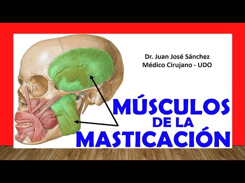
Temporalis Muscle
- The temporalis muscle originates from various bones including frontal, parietal, temporal, and sphenoid bones within the temporal fossa. It has both superficial and deep portions.
- This muscle inserts into the coronoid process of the mandible and also receives innervation from a branch of V3 (the inferior maxillary nerve), known as the temporal nerve.
- Its function complements that of the masseter by assisting in elevating and closing the mouth when clenching molars together.
Lateral Pterygoid Muscle
- Located deep to temporalis, this muscle consists of two heads: an upper head originating from greater wing crest of sphenoid bone and a lower head from lateral pterygoid plate.
Understanding the Pterygoid Muscles
Lateral Pterygoid Muscle
- The lateral pterygoid muscle inserts into the articular capsule, articular cartilage, and a pit called the Pterygoid fossa of the lower jaw. Understanding its insertion points is crucial for comprehending its function.
- Innervated by the lower maxillary nerve (third branch of the trigeminal nerve), this muscle helps in opening the mouth by moving the condyle forward during contraction.
- Unlike other muscles like temporalis and masseter that close the mouth, lateral pterygoid works synergistically with anterior digastric muscle to facilitate mouth opening and ensure proper jaw alignment during closure.
- The muscle has two portions originating from different anatomical landmarks: one from the greater crest of sphenoid and another from the external face of the lateral pterygoid plate, both inserting into the joint capsule.
- A deep view shows how these portions are positioned relative to each other as they connect to the temporomandibular joint capsule.
Medial Pterygoid Muscle
- The medial pterygoid muscle, also known as internal pterygoid, consists of superficial and deep portions that embrace part of the lateral pterygoid muscle.
- The deep portion originates from the internal face of lateral pterygoid lamina and palatine bone's pyramidal process; whereas, superficial portion also arises from this pyramidal process but is located deeper than upper jaw structures.
- Both portions insert into an area called pterygoid tuberosity on the internal face of ascending branch of lower jaw. This positioning is critical for their functional role in mastication.