🥇 PERITONEO 2/3. Epiplones, Mesos, Ligamentos y Pliegues. ¡Fácil de Entender!
Anatomy of the Peritoneum: Part 2
Overview of the Peritoneum
- Introduction to the second installment of the anatomy series on the peritoneum, emphasizing its complexity for students.
- Importance of viewing previous videos in sequence (parts one through three) before discussing retroperitoneum for comprehensive understanding.
Structures within the Abdominal Cavity
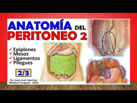
- Discussion on various structures within the abdominal cavity including omentums, mesos, ligaments, and folds.
- Definition of peritoneal folds as extensions of parietal peritoneum that can become visceral; their role in anchoring abdominal structures.
Types of Peritoneal Replicas
- Explanation of three types of peritoneal replicas: ligaments, mesos, and omentums.
- Clarification on when a fold is termed a ligament—specifically when it connects organs without significant vascular structures.
Key Ligaments Above and Below the Navel
- Identification of six key folds at navel level; only one (falciform ligament) is above while five are below.
- Description of falciform ligament's structure and function; contains round ligament which is an obliterated umbilical vein post-birth.
Detailed Examination of Lower Ligaments
- Introduction to five ligaments below the navel starting with middle umbilical ligament formed from urachus after birth.
Understanding the Anatomical Structures of the Abdominal Wall
Ligaments and Fossae of the Anterior Abdominal Wall
- The parietal peritoneum is positioned above the inferior epigastric vessels, which include both the inferior epigastric artery and vein. This positioning is crucial for understanding abdominal anatomy.
- There are various fossae formed between ligaments in the anterior abdominal wall, including:
- Internal inguinal fossa (medial inguinal fossa), located between the middle umbilical ligament and medial ligament.
- Supra-vesical fossae, situated above the bladder and pubis.
- Lateral to the external umbilical ligament are the external inguinal fossae or lateral inguinal fossae. These anatomical features are significant when discussing hernias.
- The inguinal triangle, also known as Hesselbach's triangle, is found within the middle inguinal fossa. It serves as a critical area in hernial pathology studies.
Omenta: Greater and Lesser Omentum
- Omenta are structures of visceral peritoneum that connect viscera to each other rather than to body walls. They contain important vascular pedicles with arteries and veins running through them.
- The greater omentum hangs like an apron from the greater curvature of the stomach, consisting of four layers despite some sources stating two. It has insertions at various points along the colon.
- The arrangement of leaves in the greater omentum includes:
- An anterior leaf reaching down from the stomach.
- A posterior leaf inserting into transverse colon.
- Each leaf consists of two sheets, creating a space known as transcavity.
- From an embryological perspective, this omentum derives from dorsal mesogastric tissue. Understanding its structure helps clarify its role in abdominal anatomy.
Lesser Omentum Composition
- The lesser omentum comprises two ligaments:
- The wider gastrohepatic ligament connecting liver to lesser curvature of stomach.
- The hepatoduodenal ligament linking liver to duodenum's first portion.
- Within these ligaments runs vital blood supply such as:
- Left gastric artery alongside right gastric artery forming an arch at gastric root.
Understanding the Mesentery and Omentum
Overview of Omentum and Mesentery
- The omentum has a cavity known as Winslow's hiatus, which will be discussed in detail in future segments about the peritoneum.
- A meso is defined as a vascular structure connecting the posterior abdominal wall to an organ, distinguishing it from ligaments.
Characteristics of Mesentery
- The mesentery connects the posterior abdominal wall with most of the small intestine, specifically measuring 15 centimeters from the duodenum-jejunal angle to where the ileum empties into the cecum.
- The visceral edge of the mesentery corresponds to the length of the small intestine, which can measure between 5 to 7 meters.
Dimensions and Structure
- The width of the mesentery can reach up to 20 centimeters, encompassing two leaves that contain important vascular structures like superior mesenteric artery and vein.
- The root of this mesentery extends from L1 vertebra at duodenal-jejunal angle down to iliocecal junction near right sacroiliac joint.
Transverse Mesocolon
- The transverse mesocolon supports both colic arteries (right and left), allowing them to supply blood to the transverse colon.
- It consists of two leaves: one attached to parietal peritoneum covering pancreas and another covering part of duodenum.
Ascending Mesocolon and Retroperitoneal Structures
- Ascending mesocolon is not always present; its absence means that descending colon becomes retroperitoneal.
- Mesoappendix supports appendix with appendiceal artery running along it; however, ascending/descending colon lacks such support structures.
Sigmoid Mesocolon