TIMO HISTOLOGIA
General Overview
In this section, the speaker introduces the lymphatic tissue and focuses on discussing the thymus as an organ within this tissue. The physical characteristics, variations in size, and histological features of the thymus are highlighted.
Thymus Anatomy and Function
- The thymus is a triangular organ located behind the upper portion of the sternum. Its size varies depending on age, being largest in newborns and children, decreasing in size post-puberty but persisting throughout life.
- Histologically, the thymus consists of two main regions: the cortex (outer region) and medulla (central region). The cortex contains lymphocytes at different maturation stages along with macrophages and reticular cells. In contrast, the medulla houses Hassall's corpuscles composed of concentric epithelial cells.
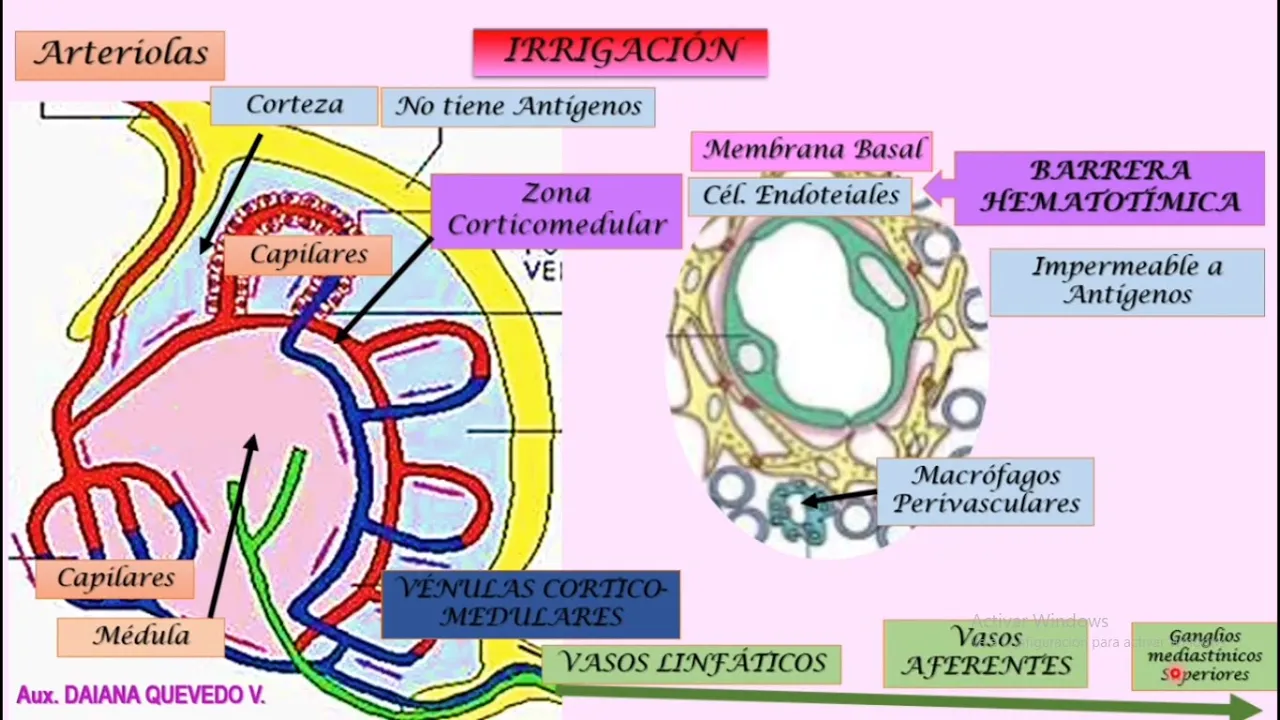
- Blood supply to the thymus involves arteries entering through septa from which capillaries branch out to irrigate both cortex and medulla. These capillaries form a barrier preventing large molecules and antigens from passing through.
Lymphatic Drainage and Cell Maturation
- Lymphatic vessels drain towards mediastinal lymph nodes after traversing septa within the thymus. These vessels are solely afferent, with no efferent vessels present within the thymic tissue.
- Regarding T lymphocyte development, precursor cells enter from blood vessels into outer cortical regions where they proliferate before migrating towards inner cortico-medullary junctions for further differentiation facilitated by epithelial cells.