5 Pediatric Orthopedic Conditions Not to Miss as a Primary Care Provider: Ellen Dean-Davis, MD
Introduction
The speaker introduces herself and her background as a pediatric orthopedic surgeon. She also provides an overview of the topics that will be covered in the presentation.
- Dr. Ellen Dean Davis is a pediatric orthopedic surgeon who grew up in Memphis, Tennessee.
- She did her orthopedic training in Paterson, New Jersey, and followed it with a pediatric orthopedic fellowship in Wilmington, Delaware.
- Dr. Davis practices at Atlantic Health System in Morristown, New Jersey.
- She discusses the three main areas of her practice: trauma and sports injuries, congenital issues like DDH and scoliosis, and complex deformity correction and neuromuscular conditions such as cerebral palsy.
Five Pediatric Orthopedic Conditions Not to Miss
The speaker discusses five pediatric orthopedic conditions that primary care practitioners should not miss.
Slipped Capital Femoral Epiphysis (SCFE)
- A 14-year-old overweight boy complains of vague left knee and thigh pain that worsens with activity.
- There is no trauma that he can remember.
- On examination, he has an antalgic gait and increased external rotation of his left foot when walking compared to the other side.
- Knee x-rays are negative.
- Next step: order hip and pelvis x-rays to check for SCFE - a transphasial fracture through the proximal femoral growth plate.
Legg-Calve-Perthes Disease (LCPD)
- A 6-year-old boy presents with a limp that has been going on for several weeks.
- He has no history of trauma or infection.
- On examination, there is limited internal rotation of his right hip with flexion and abduction.
- X-rays show a flattened femoral head with increased density.
- Next step: refer to a pediatric orthopedic surgeon for further evaluation and management.
Developmental Dysplasia of the Hip (DDH)
- A 2-month-old girl is brought in for a well-child visit.
- On examination, there is asymmetry of the gluteal folds and limited hip abduction on the left side.
- Ultrasound confirms DDH - an abnormality in the development of the hip joint that can lead to dislocation or subluxation.
- Next step: refer to a pediatric orthopedic surgeon for further evaluation and management.
Scoliosis
- A 12-year-old girl presents with back pain and her mother notices that her shoulders are uneven.
- On examination, there is a visible curve in her spine when she bends forward.
- X-rays confirm scoliosis - a lateral curvature of the spine greater than 10 degrees.
- Next step: refer to a pediatric orthopedic surgeon for further evaluation and management.
Juvenile Idiopathic Arthritis (JIA)
- An 8-year-old girl presents with morning stiffness lasting more than an hour, swelling in multiple joints, and fever.
- She has no history of trauma or infection.
- On examination, there is swelling and tenderness in multiple joints including her knees, ankles, wrists, and fingers.
- Blood tests show elevated inflammatory markers like ESR and CRP.
- Next step: refer to a pediatric rheumatologist for further evaluation and management.
Skippy: A Common Hip Disorder in Adolescents
In this section, the speaker discusses skippy, a common hip disorder in adolescents. They explain that it affects about one in every ten thousand kids and is most commonly seen in African Americans. The number one risk factor for skippy is obesity or being overweight.
Symptoms of Skippy
- Skippy presents with discomfort or vague pain throughout the groin and thigh.
- Up to 25% of children with skippy present with knee or distal thigh pain that's referred.
- Children may walk differently due to the pain, which can be noticed by parents.
- The leg will be externally rotated when walking.
Physical Examination for Skippy
- Watch the child walk up and down the hall to see if their foot turns out to the side, indicating external rotation of the hip.
- The leg will be held in external rotation, and there will be difficulty sitting in a chair due to loss of flexion.
- Obligate external rotation of the hip with hip flexion is pathognomonic for skippy.
Types of Skippy
- Abnormal load on a normal growth plate is most common and occurs due to excess weight.
- Normal load on an abnormal growth plate is less common but can occur due to endocrine disorders such as hypothyroidism or hyperparathyroidism.
Diagnosis of Skippy
- Diagnosed on plain x-ray, including both an ap and frog lateral pelvis x-ray.
- Frog lateral pelvis x-ray is where you're going to see this most easily diagnosed.
- A side-to-side comparison is necessary to diagnose subtle skippy.
Skippy: Diagnosis and Treatment
In this section, the speaker discusses the diagnosis and treatment of a condition called skippy, which is a type of slipped capital femoral epiphysis (SCFE).
Skippy Diagnosis
- X-rays are negative.
- MRI can diagnose what's called a pre-slip, which is edema, inflammation, or irritation of the growth plate that precedes the actual slip.
- Best case scenario is to catch it at this phase.
Types of Skippy
- Two types: normal load on abnormal growth plate (rare), and obesity-related (most common).
- Work up younger than expected (<10 years old), older than expected (>16 years old), and any kids who don't fit the picture of obesity.
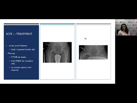
Skippy Treatment
- Put a screw across the growth plate to prevent it from getting worse.
- Post-operatively, toe touch weight-bearing for about six weeks if it's one side affected; both sides affected can bear weight for transfer purposes only.
- No contact sports until maturity.
- Encourage exercise and weight loss but not in contact sport realm.
Follow-up for Skippy
- Ask about any hip pain, groin pain, knee pain, or side pain.
- About 1 to 2 percent will continue to slip over time even after surgery.
- Contralateral slip occurs in about one-third of cases within one year from first diagnosis.
- Follow-up every three months to check if it's happening on other side and evaluate for vascular necrosis.
Genetic Component and Toe Walking
This section discusses the genetic component of obesity and family history's role in it. It also talks about toe walking, its causes, and how to diagnose it.
Genetic Component and Toe Walking
- There is no genetic component for toe walking.
- Family history doesn't play a significant role in toe walking.
- Most cases of toe walking are idiopathic or habitual.
- Underlying issues such as muscular dystrophy, cerebral palsy, or other neurologic causes can cause toe walking.
- Cerebral palsy is one of the first presentations of toe walking. Birth history is essential in diagnosing cerebral palsy.
Diagnosis of Toe Walking
- Diplegic cerebral palsy is the most likely diagnosis for a child who walks on their toes with stiffness in the Achilles tendons when dorsiflexion in the ankle.
- Idiopathic toe walkers are developmentally normal when told to walk flat. They have always walked on their toes, but there is no heel cord contracture or spasticity.
- Children with heel cord contracture have some element of spasticity. Testing the heel cord involves dorsiflexing the ankle at least 25 degrees.
Treatment for Toe Walking with Cerebral Palsy
This section discusses the treatment for toe walking with cerebral palsy.
Preventative Treatment
- The treatment for toe walking with cerebral palsy is preventative.
- Diagnosis is important to get the proper therapy needed.
- Stretching is a key part of treatment, especially as children are growing fast.
- Ankle foot orthosis may be used to supplement stretching.
Managing Heel Cord Contracture
- Heel cord contracture can be managed in various ways depending on severity.
- Serial casting, botox, and surgery are some options.
- Neurologists and physiatrists try to avoid surgery by using other methods like botox.
Toe Walking with Muscular Dystrophy
This section discusses toe walking as a presenting feature of muscular dystrophy.
Symptoms and Diagnosis
- Toe walking may be the presenting complaint or feature of someone who ends up being diagnosed with muscular dystrophy.
- Children start toe walking anywhere from two to six years old due to genetic defect causing proximal muscle weakness before distal muscles.
- Positive family history is common in these cases, and it usually affects only boys.
Treatment
- The bigger issue is making the diagnosis so that they can get started on proper therapies needed to slow disease progression.
- It's important not to take away their ability to toe walk as it may cause them to stop walking altogether.
Other Neurologic Etiologies
This section discusses other neurologic etiologies that can cause toe walking.
Symptoms and Diagnosis
- Unilateral or asymmetric toe walking is a red flag for other neurologic etiologies.
- Children with these conditions are often older than those diagnosed with muscular dystrophy.
- Abdominal reflexes, clonus, hyperreflexia, and spasticity are some symptoms to look out for.
Treatment
- An MRI of the entire spine plus or minus the brain is usually done when these conditions are suspected.
- Neurologists may be consulted to confirm if an MRI is necessary.
Monteggia Fracture-Dislocation
In this section, the speaker discusses a case of a four-year-old girl who fell off her scooter and was brought to the ER with an arm injury. The speaker explains how to diagnose a Monteggia fracture-dislocation and highlights key points to look out for.
Diagnosis of Monteggia Fracture-Dislocation
- The x-ray shows a fracture of the ulna with dislocation of the radial head.
- A Monteggia fracture-dislocation is a fracture of the ulna with dislocation of the radial head.
- A subtle bend in the ulna can cause dislocation of the radial head, which may be missed if not carefully examined.
- Forearm fractures often affect both bones rather than just one.
Pediatric Elbow Anatomy
- There are multiple ossification centers in a child's elbow that show up at different times on an x-ray, making diagnosis challenging.
- The capitellum is on the lateral side of the distal humerus and shows up around one year of age.
- The radiocapitellar line is important for diagnosing a Monteggia fracture-dislocation. It intersects with the capitellum and should be present on all views.
Plastic Deformation and Ulnar Fractures
In this section, the speaker discusses plastic deformation and ulnar fractures in children. They explain how a bowed ulna can cause the radial head to pop out of place, leading to plastic deformation. The speaker emphasizes the importance of catching this early to avoid more invasive treatments.
Plastic Deformation and Ulnar Fractures
- Plastic deformation occurs when a bowed ulna causes the radial head to pop out of place.
- Catching this early is important because it can be treated with a simple cast or minimally invasive procedures.
- Delayed treatment may require open reduction, plates, screws, and even bone cutting.
- Chronic cases may result in loss of motion or require leaving it alone.
Developmental Dysplasia of the Hip (DDH)
In this section, the speaker talks about developmental dysplasia of the hip (DDH), which is a common orthopedic condition in newborns. They discuss risk factors and emphasize the importance of screening for DDH.
Developmental Dysplasia of the Hip (DDH)
- DDH is a common orthopedic condition in newborns affecting about 100 babies.
- One in five hundred babies will have DDH as a frank dislocation.
- Screening for DDH is important, especially if there are risk factors such as family history or breech positioning during birth.
- An ultrasound should be performed at six weeks to detect any abnormalities.
Risk Factors for Hip Dysplasia
This section discusses the risk factors associated with hip dysplasia and how it is diagnosed.
Risk Factors
- Breach positioning is a solid risk factor for hip dysplasia.
- Positive family history is another significant risk factor.
- Swaddling may worsen the problem if there is known hip dysplasia or at risk for hip dysplasia.
Screening and Treatment of Hip Dysplasia
This section covers who should be screened for hip dysplasia, how it is diagnosed, and its treatment.
Who to Screen
- The American Academy of Orthopedic Surgery recommends screening if there's either breach position, family history, or clinical concern based on physical exam.
- Universal screening isn't accepted because there are too many false positives.
Diagnosis and Treatment
- Physical exam and imaging are used to diagnose hip dysplasia.
- Ultrasound is used if the baby is under four months old, while an x-ray is used if they're over six months old.
- Treatment involves using a pelvic harness that has some risks such as brachial plexus palsy or femoral nerve palsy.
- If there's a positive Barlow and Ortolani maneuver, treatment starts before getting an ultrasound.
When to Send for Ultrasound
This section discusses when to send babies for ultrasound testing to confirm diagnosis of hip dysplasia.
Timing of Screening Exam
- The screening exam should be done between four and six weeks of age because maternal estrogen makes everything super loose in newborns.
- Doing the ultrasound too early has a high false positive rate so waiting until six weeks is better.
Ultrasound vs X-ray
- Ultrasound is preferred for babies between four to six months old because it's a dynamic study that provides more information.
- After six months, an x-ray is used instead of ultrasound.
Physical Exam for Hip Dysplasia
This section covers the physical exam used to diagnose hip dysplasia.
Barlow and Ortolani Maneuver
- The Barlow maneuver involves dislocating the hip while the Ortolani maneuver involves abducting the hips and reducing or putting it right back in place.
- These maneuvers are not painful for babies and are used to diagnose hip dysplasia.
Developmental Dysplasia of the Hip (DDH)
In this section, the speaker discusses developmental dysplasia of the hip (DDH), its diagnosis, and treatment.
Diagnosis
- DDH is diagnosed by looking for gluteal fold asymmetry.
- After three months, hip abduction is limited and can cause walking difficulties.
- A hip click is not a reliable indicator of DDH.
Treatment
- The Pavlik harness is used to treat DDH in infants. It keeps the femoral head pointed towards the acetabulum and allows for motion through the leg.
- The harness should be worn until there is improvement, which usually takes 6 to 12 weeks depending on severity.
- If a baby fails Pavlik treatment, a more rigid brace can be used before resorting to a spica cast.
- Spica casting is a last resort and requires full-time care for at least three months.
Finger Hematoma
In this section, the speaker discusses an eight-year-old boy who presents with a finger hematoma after slamming his finger in a car door.
Diagnosis
- No timestamps available for this section.
Treatment
- No timestamps available for this section.
Seymour Fracture and Other Pediatric Orthopedic Emergencies
In this talk, the speaker discusses pediatric orthopedic emergencies such as seymour fracture, developmental dysplasia of the hip (DDH), and toe walking. The focus is on recognizing these conditions and providing appropriate management to prevent complications.
Seymour Fracture
- A seymour fracture is a distal phalangeal fracture with an associated nail bed injury that can occur in fingers or toes.
- It is often caused by direct trauma or crush injuries.
- Symptoms include nail bed laceration, sublingual hematoma, and bulbous appearance at the base of the nail.
- X-rays may appear normal but can show widening of the growth plate or flexion deformity.
- If left untreated, it can result in chronic osteomyelitis.
- Urgent evaluation is necessary to avoid complications.
- Treatment involves taking up the nail, cleaning out debris, stabilizing the fracture with a splint or pin, and administering antibiotics.
Developmental Dysplasia of the Hip (DDH)
- DDH is a condition where there is abnormal development of the hip joint that can lead to dislocation or subluxation.
- Risk factors include female sex, breech presentation at birth, family history of DDH, and oligohydramnios.
- Screening involves physical exam using techniques such as Barlow's maneuver and Ortolani's test.
- Early detection and treatment are important to prevent long-term complications such as arthritis and gait abnormalities.
Toe Walking
- Toe walking is a common condition in children where they walk on their toes instead of their heels.
- It can be idiopathic or due to underlying neurological or musculoskeletal conditions.
- Diagnosis involves taking a thorough history and performing a physical exam to rule out underlying conditions.
- Treatment depends on the underlying cause and may involve physical therapy, orthotics, or surgery.
Skipped Content
- There is no relevant content skipped in this transcript.
Q&A Session
In this section, the speaker answers questions from the audience.
Ultrasound for Premature Infants
- Premature infants can be ultrasounded at six weeks after birth if there is high suspicion because maternal estrogen has cleared the system and false positives are less likely.
Asymmetric Toe Walking
- Asymmetric toe walking requires workup to determine the underlying cause, which could be due to an underlying neurologic or genetic condition.
Subungual Hematoma and X-rays
- Any subungual hematoma with active bleeding, nail bed laceration around it, or elevation of tissue proximal to the nail should have an x-ray.
Skippy Diagnosis for Obese Patients
- If clinical suspicion is very high, obese patients with hip and knee pain should be sent straight to the emergency room instead of getting an x-ray first.
Conclusion
- The speaker thanks attendees for participating in the event and offers his contact information for further questions.
Turn any video into a summary like this
YouTube links, meetings, lectures. With transcripts, search, and chat.