Trachea, Bronchial Tree and Alveolar Tree (Parts, Structures and Walls) - Anatomy
Anatomy of the Respiratory System: Trachea and Bronchi
Overview of the Respiratory System
- The respiratory system includes organs involved in breathing: Nose, nasal cavity, Pharynx, Larynx, Trachea, Bronchi, and Lungs.
- Previous videos covered the anatomy of the nasal cavity and larynx.
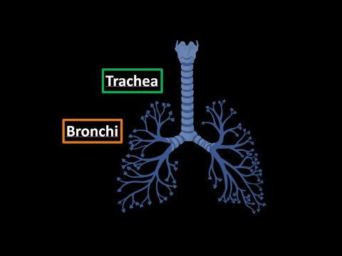
Anatomy of the Trachea
- The trachea is also known as the windpipe; it directs air to and from the lungs. It measures 9 to 15 cm in length and has a diameter of about 2 to 2.5 cm.
- The trachea extends from the sixth or seventh cervical vertebra down to the fourth or fifth thoracic vertebrae.
Structural Components
- The tracheal wall consists of horseshoe-shaped cartilages (tracheal cartilages) that provide structural support without covering the back side completely. These are connected by annular ligaments containing strong fibers.
- A fibromuscular membrane connects these cartilages at their dorsal edges, forming what is known as the membranous part of the trachea. This structure includes tracheal muscles and connective tissue for flexibility and strength.
Layers of Tracheal Wall
- The innermost layer is called tunica mucosa, which contains lymphoid nodules that combat microorganisms and glands that lubricate the respiratory tract surface. Coughing helps expel trapped irritants.
- Following this is tela submucosa with loose connective tissue and blood vessels; then comes cartilage followed by tunica adventitia—dense connective tissue providing external protection for the trachea.
Bifurcation into Bronchi
- At its lower end, where it splits into bronchi (tracheal bifurcation), there’s an elevation called Carina of Trachea—a ridge formed by last cartilage before branching into bronchi while being located anteriorly to esophagus.
Anatomy of Bronchi
- The right primary bronchus (about 2.5 cm long) is shorter, wider, more vertical compared to left primary bronchus (about 5 cm long). They split at approximately fourth or fifth thoracic vertebrae where trachea ends.
Bronchial Tree Structure and Function
Overview of Bronchi Division
- The primary bronchus divides into smaller bronchi based on lung lobes: three lobar bronchi for the right lung and two for the left.
- Each lobar bronchus further divides into segmental bronchi, with 10 segments in the right lung and 8 in the left.
Detailed Anatomy of Bronchi
- The primary bronchus enters through the hilum, branching into superior, middle, and inferior lobar bronchi on the right; only superior and inferior on the left.
- Each lobe is anatomically divided into segments; this division is crucial for surgical procedures involving tumor removal or other interventions.
Importance of Segmental Division
- Understanding segmental division aids in surgical resection without damaging surrounding lung tissue.
- The bronchial tree consists of main bronchi that divide into lobar (2 or 3 per side), then segmental (10 right, 8 left), continuing to split until reaching terminal bronchioles.
Transition to Alveolar Tree
- Terminal bronchioles are approximately 0.5–1 mm in diameter; they mark the end of the bronchial tree before transitioning to alveolar structures.
- Respiratory epithelium lines the bronchial tree, which protects airways by trapping irritants with cilia.
Gas Exchange Mechanism
- At terminal bronchioles, respiratory epithelium changes to cuboidal cells as it transitions to alveolar structures where gas exchange occurs.
- Alveoli increase deeper within lungs; secondary and tertiary bronchioles contain more alveoli for efficient gas exchange.
Histological Differences in Airway Structures
Epithelium Changes Along Bronchial Tree
- The inner lining of the trachea features respiratory epithelium; as one moves downwards through bronchi and bronchioles, epithelial types change from ciliated to cuboidal/squamous cells.
Structural Layers Comparison
- Trachea's structure includes tunica mucosa lined with respiratory epithelium, a submucosal layer with loose connective tissue, cartilage rings, smooth muscle at its back, and an outer protective layer called tunica adventitia.
Bronchus vs. Bronchiole Structure
- In contrast to trachea structure:
- Bronchus retains tunica mucosa but has a fibro-musculo-cartilaginous layer instead of distinct cartilage rings.
- Cartilage diminishes as it transitions towards smaller airways like bronchioles.