THE CYTOSKELETON - MICROTUBULES, INTERMEDIATE FILAMENTS, MICROFILAMENTS
Cytoskeleton Structure and Function
Overview of Cytoskeletal Filaments
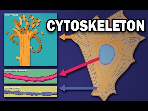
- The cytoskeleton in animal cells consists of three types of filaments: microtubules, intermediate filaments, and microfilaments. These structures provide cellular support, facilitate movement, and assist in intracellular transport.
Microtubules
- Microtubules organize organelle positions and direct intracellular transport. They are crucial for maintaining cell structure.
Intermediate Filaments
- Intermediate filaments are rope-like fibers that provide mechanical strength and connect epithelial cells through a network of cables along the nuclear envelope.
Microfilaments (Actin Filaments)
- Microfilaments are concentrated beneath the plasma membrane (cortex), controlling cell shape and locomotion. They form projections like microvilli, lamellopodia, and filipodia to aid in movement over solid surfaces.
Dynamic Nature of the Cytoskeleton
Assembly and Disassembly
- The cytoskeleton is dynamic; its components can rapidly assemble or disassemble based on cellular needs due to weak non-covalent linkages between subunits.
Nucleation Process
- Nucleation is the rate-limiting step for polymer formation. Initial aggregates are unstable but become more stable as multiple protofilaments associate laterally.
Cellular Requirements for Cytoskeletal Structures
Rapid Rearrangement vs Stability
- Different cells have varying requirements: white blood cells need rapid cytoskeletal rearrangement for movement, while neurons require stable structures for functionality.
Epithelial Cell Polarity
- In epithelial tissues, the cytoskeleton maintains polarity throughout a cell's life cycle by organizing specialized surface protrusions that enhance nutrient transfer.
Microfilament Details
Structure and Dynamics
- Microfilaments consist of actin protein arranged in double helices with diameters of 6 nm. They form various networks that exhibit properties similar to semi-solid gels.
ATP Binding and Treadmilling
- Actin monomers bind to ATP during polymer growth; hydrolysis to ADP increases dissociation likelihood from filament ends. Treadmilling occurs when one end grows while the other shrinks at equilibrium concentrations.
Nucleation Mechanisms
ARP Complex Role
- The ARP complex promotes nucleation by attaching to existing actin filaments, facilitating rapid growth from their plus ends into branched networks at angles around 70°.
Accessory Proteins Impacting Actin Dynamics
Capping Proteins and Depolymerization
- Capping proteins stabilize actin filaments by preventing growth or shrinkage; cofilin promotes depolymerization of older filaments while allowing forward movement of the network despite stationary individual filaments.
Intermediate Filament Characteristics
Composition and Functionality
- Intermediate filaments measure 10 nm in diameter, composed of fibrous proteins that maintain cell shape under tension. They anchor organelles like the nuclear lamina within epithelial tissues through desmosomes.
Microtube Structure & Properties
Composition
- Microtuples are hollow tubes made from tubulin dimers (alpha & beta). Thirteen protofilament arrangements create a rigid structure with diameters ranging from 25 nm externally to 15 nm internally.
Polar Nature
Microtubule Dynamics and Organization
ATP and GTP Hydrolysis in Filament Growth
- ATP is hydrolyzed to ADP shortly after actin molecules are added to a growing filament, while microtubules have GTP tightly bound to tubulin heterodimers.
- Upon binding of the dimer into a growing microtubule, GTP is soon hydrolyzed to GDP, which decreases the affinity of the subunit for lateral protofilaments.
Dynamic Instability of Microtubules
- Dynamic instability arises from differences between the two polar ends of microtubules; if subunit addition exceeds GTP hydrolysis, a GTP cap forms.
- A microtubule without a GTP cap depolymerizes approximately 100 times faster than one with it. The loss of this cap leads to rapid shrinkage known as catastrophe.
- If sufficient GTP-bound subunits are added quickly enough during shrinkage, an event called rescue can occur, forming a new cap.
Microtubule Nucleation and Organization
- Microtubules are primarily nucleated near the nucleus and organized by microtubule organizing centers (MTOCs), such as centrosomes containing centrioles arranged at right angles.
- Centrosomes serve as spindle poles during mitosis and meiosis, facilitating chromosome separation through rapid cytoskeletal reorganization post-replication.
Structure of Cilia and Flagella
- Basal bodies also function as MTOCs found in cilia and flagella, which share a common cross-section featuring a 9+2 structure: nine doublets of microtubules surrounding two single microtubules.