🥇 Articulación TEMPOROMANDIBULAR. Fácil, Rápida y Sencilla
Articulación Temporomandibular: Anatomía y Función
Introducción a la Articulación Temporomandibular
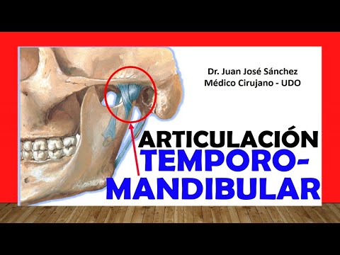
- La articulación temporomandibular (ATM) se forma entre el maxilar inferior (mandíbula) y el hueso temporal del cráneo.
- Se menciona que "articulación temporo maxilar" es un sinónimo de ATM, destacando su importancia en la anatomía facial.
Estructura de la Articulación
- La ATM incluye el cóndilo del maxilar inferior que se articula con la fosa condílea del hueso temporal.
- El hueso temporal es crucial en esta articulación, junto con el esfenoides, que también juega un papel importante en la base del cráneo.
Disco Articular y Subarticulaciones
- Un disco fibrocartilaginoso irregular separa las dos subarticulaciones dentro de la ATM, permitiendo movimientos específicos.
- La parte superior permite deslizamientos (movimientos de protección, retracción y lateralización), mientras que la parte inferior funciona como una bisagra para abrir y cerrar la boca.
Relaciones Anatómicas Importantes
- Lateralmente, la ATM está situada anterior al pabellón auricular y detrás del arco cigomático; es superficial y accesible al tacto.
- Medialmente, se relaciona con estructuras como la espina del esfenoides y los agujeros espinoso y oval; también tiene relación con partes de la faringe.
Cápsula Articular
- La cápsula articular fibrosa recubre toda la articulación sinovial e inserta en los bordes del cóndilo y cavidad condílea del hueso temporal.
Understanding the Temporomandibular Joint (TMJ)
Anatomy and Function of the TMJ
- The contraction of specific muscles allows for the forward movement of the maxillary condyle, initiating mouth opening and protrusion. This highlights the importance of muscle initiation at the level of the articular capsule.
- The articular disc is irregularly shaped, with a concave-convex upper surface and a completely concave lower surface that adapts to the inferior maxillary condyle. This disc plays a crucial role in joint function during mouth movements.
- The temporomandibular joint (TMJ) is unique as it can luxate physiologically, meaning it can lose contact between its surfaces without being pathological. Ligaments help prevent pathological luxation by stabilizing the joint.
Ligaments Supporting TMJ Stability
- Three key ligaments reinforce the TMJ:
- External ligament: Also known as temporomandibular or tempormaxilar ligament; connects from the temporal bone's articular tubercle to the external face of the ascending ramus of the mandible.
- Sphenomandibular ligament: Extends from sphenoid bone to lingula, covering a groove on the ascending ramus of mandible; important for medial relations involving internal maxillary artery and auriculotemporal nerve.
- Styloid-mandibular ligament: Connects from styloid process of temporal bone to angle and posterior border of ascending ramus; provides additional support to TMJ structure.
Movements Facilitated by TMJ
- Opening/Closing Movements:
- Mouth opening (depression) is facilitated by lateral pterygoid muscle and infrahyoid muscles including digastric, mylohyoid, and geniohyoid; gravity also assists this action.
- Closing (elevation) involves temporal muscle, masseter muscle, and medial pterygoid muscle working together for effective jaw closure.
- Protrusion/Retrusion Movements:
- Protrusion (anterior sliding) is achieved through medial pterygoid, lateral pterygoid, and masseter muscles.
- Retrusion (posterior sliding) relies on posterior fibers of temporal muscle for returning jaw backward into position.
- Lateralization Movements:
- Lateral movement requires contraction of left-side temporal and masseter muscles along with contralateral lateral pterygoids; similar mechanics apply when moving towards opposite side using corresponding muscles on that side.
This structured overview captures essential insights about TMJ anatomy, supporting ligaments, and functional movements based on provided timestamps from your transcript while ensuring clarity in understanding complex concepts related to this critical joint system.