micro ufsb03 parteC
Overview of Cell Wall Structure in Bacteria
Function and Importance of the Cell Wall
- The cell wall serves as a structural boundary, maintaining the shape of bacteria and providing protection from environmental factors.
- It plays a crucial role during cell division by ensuring that genetic material is duplicated and properly segregated into two daughter cells.
Gram Staining Technique
- The discussion references the historical development of the Gram staining technique by Hans Christian Gram in the late 1800s, which differentiates bacterial types based on their cell wall composition.
- This method allows for the classification of bacteria into two main groups: Gram-positive and Gram-negative, based on their cell wall thickness and lipid content.
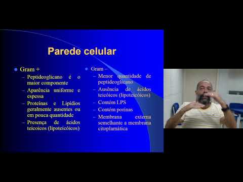
Characteristics of Gram-positive vs. Gram-negative Bacteria
- Gram-positive bacteria have a thick peptidoglycan layer with minimal lipids, making them structurally robust. This layer is essential for retaining crystal violet dye during staining processes.
- Gram-negative bacteria, in contrast, possess a thinner peptidoglycan layer surrounded by an outer membrane rich in lipopolysaccharides (LPS), which can affect permeability to antibiotics.
Implications for Antibiotic Treatment
- Understanding these differences is critical for antibiotic development; specific antibiotics target either Gram-positive or Gram-negative bacteria based on their unique cell wall structures. For instance, some antibiotics are designed to penetrate only one type effectively while others are broad-spectrum but less potent against both types.
- The presence or absence of certain components like LPS influences how effective an antibiotic will be against different bacterial strains, highlighting the importance of accurate bacterial identification through techniques like Gram staining.
Summary of Key Structural Differences
- In summary:
- Gram-positive: Thick peptidoglycan layer; few lipids; retains dye well.
- Gram-negative: Thin peptidoglycan layer; significant lipid presence; more complex structure affecting permeability and antibiotic susceptibility.
How to Differentiate Between Gram-Positive and Gram-Negative Bacteria
Overview of the Staining Process
- The process involves using oil immersion at a 1000x magnification to observe bacterial cells. If the cells appear blue, they are gram-positive; if red, they are gram-negative.
- This staining technique allows for differentiation between the structures of gram-positive and gram-negative bacteria through specific color changes during observation.
Steps in the Procedure
- The entire procedure can take approximately 12 to 15 minutes, with an additional minute required for final observations under a microscope.
- After applying the stain, it is essential to rinse and prepare the slide properly before examining it with a high-powered lens for accurate results.