Las Meninges: Duramadre, Aracnoides y Piamadre
Understanding the Meninges
Introduction to the Meninges
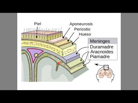
- The video introduces the topic of meninges, explaining their layers and functions. It begins by discussing the scalp, which is distinct from other skin types due to its thickness and vascularization.
Layers Covering the Head
- The scalp is described as thick and highly vascularized, leading to significant bleeding when injured. Below the scalp lies the epicranial aponeurosis (galea), a muscle that extends around the head.
Structure Beneath the Scalp
- Underneath the epicranial aponeurosis is the periosteum, which covers bones. There are two types: external periosteum in contact with galea and internal periosteum adjacent to dura mater.
Overview of Meninges
- The meninges consist of three layers: dura mater (outermost), arachnoid mater (middle), and pia mater (innermost). They protect both the brain and spinal cord.
Dura Mater Characteristics
- Dura mater is noted for being thickest and strongest among meninges. It has two layers: a periosteal layer against bone and a meningeal layer next to arachnoid mater.
Dural Venous Sinuses
Importance of Dural Sinuses
- The separation between dura mater's two layers forms venous sinuses that collect blood from cerebral veins. This process also involves cerebrospinal fluid (CSF).
Blood Supply to Dura Mater
- Understanding arterial supply to dura mater is crucial since trauma can damage these arteries, causing complications. The middle meningeal artery primarily supplies this layer.
Meningeal Layers Comparison
Arachnoid Mater vs Pia Mater
- Arachnoid mater features thin extensions resembling spider legs that create a subarachnoid space filled with CSF, essential for cushioning brain tissue.
Subarachnoid Space Functionality
Understanding the Protective Layers of the Brain
The Meninges and Cerebrospinal Fluid
- The pia mater is a very thin layer of the meninges, providing visceral protection to the brain. It is more delicate compared to the dura mater, which offers primary protection.
- The brain floats in cerebrospinal fluid (CSF), which cushions it from impacts and prevents damage from bony structures at the base of the skull.
- The spinal cord has a similar protective structure as the brain, with layers including dura mater, arachnoid, subarachnoid space, and pia mater surrounding it.
Accumulation of Blood in Head Trauma
- Caput succedaneum refers to swelling or blood accumulation under the scalp, common in newborns after difficult births; it appears soft and can be alarming but is usually harmless.
- A subgaleal hematoma occurs between the galea aponeurotica (epicranial aponeurosis) and periosteum when blood collects there; this condition can also arise from trauma.
Types of Hematomas
- A cephalohematoma forms between periosteum and skull bone; it's harder than caput succedaneum and typically results from head injuries in children.
- An epidural hematoma occurs above the dura mater due to arterial rupture; it develops quickly and poses significant danger due to rapid bleeding.
- A subdural hematoma forms beneath the dura mater from venous bleeding; it grows slower than an epidural hematoma but still requires careful monitoring.
Critical Conditions Related to Blood Accumulation
- Subarachnoid hemorrhage is extremely dangerous and requires immediate medical attention.